A importância dos níveis de vitamina D nas doenças autoimunes

A importância dos níveis de vitamina D nas doenças autoimunes

Revista Brasileira de Reumatologia

Print version ISSN 0482-5004

Rev. Bras. Reumatol. vol.50 no.1 São Paulo Jan./Feb. 2010

http://dx.doi.org/10.1590/S0482-50042010000100007

ARTIGO DE REVISÃO

 http://www.scielo.br/scielo.php?pid=S0482-50042010000100007&script=sci_arttext&tlng=eshttp://www.scielo.br/scielo.php?pid=S0482-50042010000100007&script=sci_arttext&tlng=es

A importância dos níveis de vitamina D nas doenças autoimunes

Cláudia Diniz Lopes MarquesI, Andréa Tavares DantasII, Thiago Sotero FragosoIII, Ângela Luzia Branco Pinto DuarteIV

IReumatologista, Doutora em Saúde Pública e Tutora da Escola Pernambucana de Medicina – FBV/IMIP
IIEx-residente do Serviço de Reumatologia do HC-UFPE e aluna do Curso de Mestrado do Programa de Pós-Graduação em Ciências da Saúde da UFPE
IIIResidente de Reumatologia Pediátrica do HC-UFPE e aluno do Curso de Mestrado do Programa de Pós-Graduação em Ciências da Saúde da UFPE
IVProfessora Titular e Chefe do Serviço de Reumatologia do HC-UFPE

Endereço para correspondência


RESUMO

Além do seu papel na homeostase do cálcio, acredita-se que a forma ativa da vitamina D apresenta efeitos imunomoduladores sobre as células do sistema imunológico, sobretudo linfócitos T, bem como na produção e na ação de diversas citocinas. A interação da vitamina D com o sistema imunológico vem sendo alvo de um número crescente de publicações nos últimos anos. Estudos atuais têm relacionado a deficiência de vitamina D com várias doenças autoimunes, comodiabetes mellitus insulino-dependente (DMID), esclerose múltipla (EM), doença inflamatória intestinal (DII), lúpus eritematoso sistêmico (LES) e artrite reumatoide (AR). O artigo faz uma revisão da fisiologia e do papel imunomodulador da vitamina D, enfatizando sua participação nas doenças reumatológicas, como o lúpus e a artrite reumatoide.

Palavras-chave: vitamina D, sistema imunológico, doenças autoimunes, lúpus eritematoso sistêmico, artrite reumatoide.


INTRODUÇÃO

A vitamina D e seus pró-hormônios têm sido alvo de um número crescente de pesquisas nos últimos anos, demonstrando sua função além do metabolismo do cálcio e da formação óssea, incluindo sua interação com o sistema imunológico, o que não é uma surpresa, tendo em vista a expressão do receptor de vitamina D em uma ampla variedade de tecidos corporais como cérebro, coração, pele, intestino, gônadas, próstata, mamas e células imunológicas, além de ossos, rins e paratireoides.1

Estudos atuais têm relacionado a deficiência de vitamina D com várias doenças autoimunes, incluindo diabetes melito insulino-dependente (DMID), esclerose múltipla (EM), doença inflamatória intestinal (DII), lúpus eritematoso sistêmico (LES) e artrite reumatoide (AR).1-4 Diante dessas associações, sugere-se que a vitamina D seja um fator extrínseco capaz de afetar a prevalência de doenças autoimunes.5

A vitamina D parece interagir com o sistema imunológico através de sua ação sobre a regulação e a diferenciação de células como linfócitos, macrófagos e células natural killer (NK), além de interferir na produção de citocinas in vivo in vitro. Entre os efeitos imunomoduladores demonstrados destacam-se: diminuição da produção de interleucina-2 (IL-2), do interferon-gama (INFγ) e do fator de necrose tumoral (TNF); inibição da expressão de IL-6 e inibição da secreção e produção de autoanticorpos pelos linfócitos B.6,7

FISIOLOGIA DA VITAMINA D

A vitamina D, ou colecalciferol, é um hormônio esteroide, cuja principal função consiste na regulação da homeostase do cálcio, formação e reabsorção óssea, através da sua interação com as paratireoides, os rins e os intestinos.8

A principal fonte da vitamina D é representada pela formação endógena nos tecidos cutâneos após a exposição à radiação ultravioleta B.8-10 Uma fonte alternativa e menos eficaz de vitamina D é a dieta, responsável por apenas 20% das necessidades corporais, mas que assume um papel de maior importância em idosos, pessoas institucionalizadas e habitantes de climas temperados.9

Quando exposto à radiação ultravioleta, o precursor cutâneo da vitamina D, o 7-desidrocolesterol, sofre uma clivagem fotoquímica originando a pré-vitamina D3. Essa molécula termolábil, em um período de 48 horas, sofre um rearranjo molecular dependente da temperatura, o que resulta na formação da vitamina D3 (colecalciferol). A pré-vitamina D3 também pode sofrer um processo de isomerização originando produtos biologicamente inativos (luminosterol e taquisterol) e esse mecanismo é importante para evitar a superprodução de vitamina D após períodos de prolongada exposição ao sol. O grau de pigmentação da pele é outro fator limitante para a produção de vitamina D, uma vez que peles negras apresentam limitação à penetração de raios ultravioleta.9

No sangue, a vitamina D circula ligada principalmente a uma proteína ligadora de vitamina D, embora uma pequena fração esteja ligada à albumina.No fígado, sofre hidroxilação, mediada por uma enzima citocromo P450-like, e é convertida em 25-hidroxivitamina D [25(OH)D] que representa a forma circulante em maior quantidade, porém biologicamente inerte.8,9 A etapa de hidroxilação hepática é pouco regulada, de forma que os níveis sanguíneos de 25(OH)D refletem a quantidade de vitamina D que entra na circulação, sendo proporcional à quantidade de vitamina D ingerida e produzida na pele.9,10

A etapa final da produção do hormônio é a hidroxilação adicional que acontece nas células do túbulo contorcido proximal no rim, originando a 1,25 desidroxivitamina D [1,25(OH)2D3], sua forma biologicamente ativa.8,9

Reconhece-se, atualmente, a existência da hidroxilação extrarrenal da vitamina D, originando a vitamina que agiria de maneira autócrina e parácrina, com funções de inibição da proliferação celular, promoção da diferenciação celular e regulação imunológica. A regulação da atividade da 1-a-hidroxilase renal é dependente da ingestão de cálcio e fosfato, dos níveis circulantes dos metabólitos da 1,25(OH)2De do paratormônio (PTH). Por outro lado, a regulação da hidroxilase extrarrenal é determinada por fatores locais, como a produção de citocinas e fatores de crescimento, e pelos níveis de 25(OH)D, tornando essa via mais sensível à deficiência de vitamina D.10

A principal função da vitamina D consiste no aumento da absorção intestinal de cálcio, participando da estimulação do transporte ativo desse íon nos enterócitos.9,11Atua, também, na mobilização do cálcio a partir do osso, na presença do PTH, e aumenta a reabsorção renal de cálcio no túbulo distal.12 A deficiência prolongada de vitamina D provoca raquitismo e osteomalacia e, em adultos, quando associada à osteoporose, leva a um risco aumentado de fraturas.13

Outras ações da vitamina D regulando positivamente a formação de osso incluem: inibição da síntese de colágeno tipo 1; indução da síntese de osteocalcina; promoção da diferenciação, in vitro, de precursores celulares monócitos-macrófagos em osteoclastos. Além disso, estimula a produção do ligante RANK (RANK-L), o que resulta em um efeito que facilita a maturação dos precursores de osteoclastos para osteoclastos, que, por sua vez, mobilizam os depósitos de cálcio do esqueleto, para manter a homeostase do cálcio.8,9 A vitamina D exerce suas funções biológicas através da sua ligação a receptores nucleares, os receptores para vitamina D (RVD), que regulam a transcrição do DNA em RNA, semelhante aos receptores para esteroides, hormônios tireoidianos e retinoides.9,11 Esses receptores são expressos por vários tipos de células, incluindo epitélio do intestino delgado e tubular renal, osteoblastos, osteoclastos, células hematopoiéticas, linfócitos, células epidérmicas, células pancreáticas, miócitos e neurônios.11

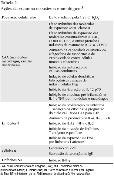
Mais recentemente, têm sido evidenciadas as ações não calcêmicas da vitamina D, mediadas pelo RVD, como proliferação e diferenciação celular, além de imunomodulação. O RVD é amplamente expresso na maioria das células imunológicas, incluindo monócitos, macrófagos, células dendríticas, células NK e linfócitos T e B.14 No entanto, sua maior concentração está em células imunológicas imaturas no timo e nos linfócitos CD8 maduros, independentemente do seu estado de ativação.8,12 O resumo dos principais efeitos da vitamina D no sistema imunológico pode ser observado na Tabela 1.

 1TS

MENSURAÇÃO DA VITAMINA D SÉRICA

Embora a forma ativa da vitamina D seja a 1,25OH2D3, esta não deve ser utilizada para avaliar sua concentração sérica, uma vez que sua meia-vida é de apenas 4 h e sua concentração é 1.000 vezes menor do que a de 25(OH)D. Além disso, no caso de deficiência de vitamina D, existe um aumento compensatório na secreção do PTH, o que estimula o rim a produzir mais a 1,25OH2D3. Desse modo, quando ocorre deficiência de vitamina D e queda dos níveis de 25(OH)D, as concentrações de 1,25OH2Dse mantêm dentro dos níveis normais e, em alguns casos, até mesmo mais elevadas.9,15

Para quantificar se existem níveis adequados de vitamina D, deve ser dosada a concentração de 25(OH)D, que representa sua forma circulante em maior quantidade, com meia-vida de cerca de duas semanas.9

Não existe consenso sobre a concentração sérica ideal de vitamina D. A maioria dos especialistas concorda que o nível de vitamina D deva ser mantido em uma faixa que não induza aumento dos níveis de PTH.10,15 Os valores normais nos diversos ensaios comerciais existentes variam de 25 a 37,5 nmol/L (10 a 15 ng/mL) a 137,5 a 162,5 nmol/L (55 a 65 ng/mL)13. Revisões recentes sugerem que a concentração ideal seja de 50 a 80 nmol/L enquanto outros sugerem valores entre 75 e 125 nmol/L.16Usando a elevação do PTH como biomarcador refletindo baixos níveis fisiológicos de vitamina D, a deficiência deve ser definida como concentração sérica inferior a 32 ng/mL (80 nmol/L).16

O nível ideal de vitamina D necessário para garantir o bom funcionamento do sistema imunológico ainda não está definido. Provavelmente, esse valor deve ser diferente daquele necessário para prevenir a deficiência de vitamina D ou manter a homeostase do cálcio.5

AÇÃO DA VITAMINA D SOBRE O SISTEMA IMUNOLÓGICO

Com base na produção ectópica de vitamina D em células do sistema imunológico e na presença de RVD em tecidos não relacionados com a fisiologia óssea, as propriedades imunorreguladoras da vitamina D têm sido cada vez mais bem caracterizadas.17 Estudos epidemiológicos mostram que a deficiência de vitamina D poderia estar associada a risco aumentado de neoplasia de cólon e próstata, doença cardiovascular e infecções.13,15,17,18

Vários mecanismos têm sido propostos para explicar a participação da vitamina D na fisiologia do sistema imunológico, conforme pode ser observado na Tabela 1.

Dentre as principais funções da vitamina D no sistema imunológico podemos destacar: regulação da diferenciação e ativação de linfócitos CD4;5,11,19 aumento do número e função das células T reguladoras (Treg);11 inibição in vitro da diferenciação de monócitos em células dendríticas;8,11 diminuição da produção das citocinas interferon-g, IL-2 e TNF-a, a partir de células Th1 e estímulo da função células Th2helper;5,8,11,17 inibição da produção de IL-17 a partir de células Th1720 e estimulação de células T NK in vivo e in vitro.21

VITAMINA D E DOENÇAS AUTOIMUNES

De maneira geral, o efeito da vitamina D no sistema imunológico se traduz em aumento da imunidade inata associado a uma regulação multifacetada da imunidade adquirida.22 Tem sido demonstrada uma relação entre a deficiência de vitamina D e a prevalência de algumas doenças autoimunes como DMID, EM, AR, LES e DII.5,13

Sugere-se que a vitamina D e seus análogos não só previnam o desenvolvimento de doenças autoimunes como também poderiam ser utilizados no seu tratamento.11 A suplementação de vitamina D tem-se mostrado terapeuticamente efetiva em vários modelos animais experimentais, como encefalomielite alérgica, artrite induzida por colágeno, diabetes melito tipo 1, doença inflamatória intestinal, tireoidite autoimune e LES.8

Baixos níveis séricos de vitamina D podem, ainda, estar relacionados com outros fatores como diminuição da capacidade física, menor exposição ao sol, maior frequência de polimorfismos nos genes do RVD, efeito colateral de medicamentos, além de fatores nutricionais.10,11

Artrite reumatoide

A AR é uma doença imunomediada, com fisiopatologia bastante complexa. Acredita-se que o evento inicial seja provavelmente a ativação de células T dependente de antígenos, desencadeando uma resposta imunológica essencialmente do tipo Th1. Essa ativação leva a múltiplos efeitos, incluindo ativação e proliferação de células endoteliais e sinoviais, recrutamento e ativação de células pró-inflamatórias, secreção de citocinas e proteases a partir de macrófagos e células sinoviais fibroblastos-like e produção de autoanticorpos.23

Como enfatizado anteriormente, a deficiência de vitamina D está associada à exacerbação da resposta imunológica Th1. Dessa forma, nos últimos anos, a participação da vitamina D na patogênese, atividade e tratamento da AR tem sido aventada com base nos resultados e nas observações de estudos clínicos e experimentais.5

O fundamento para relacionar deficiência de vitamina D e AR se baseia em dois argumentos: existem evidências de que ocorre deficiência de vitamina D em pacientes com AR e a presença de 1,25(OH)2De do RVD em macrófagos, condrócitos e sinoviócitos nas articulações destes pacientes.14,24

A relação entre polimorfismos do gene do RVD e o início e atividade da AR foi demonstrada em um estudo no qual pacientes com genótipos BB ou Bb para RVD tiveram maiores índices no HAQ, velocidade de hemossedimentação (VSH), uso cumulativo de corticosteroides e número de medicamentos antirreumáticos modificadores da doença (DMARD) em comparação com pacientes com o genótipo BB.25

Em modelos de artrite induzida por colágeno, a suplementação dietética ou administração oral de vitamina D preveniu o desenvolvimento de artrite ou retardou a sua progressão.1,26 Da mesma forma, um estudo com 29.368 mulheres mostrou que maior ingestão de vitamina D foi inversamente associada ao risco de desenvolvimento da AR.27 No entanto, outra grande coorte prospectiva, que avaliou 186.389 mulheres no período de 1980 a 2002, não encontrou associação entre ingestão aumentada de vitamina D e risco de desenvolver AR ou LES.28Corroborando esse resultado, um estudo com 79 doadores de sangue, avaliando a dosagem sérica de vitamina D, não mostrou diferenças entre os níveis basais de vitamina D nos pacientes que, posteriormente, desenvolveram AR em comparação com os controles.29 Estes achados demonstram que o assunto ainda é bastante controverso, não havendo consenso entre os autores sobre a real relação entre vitamina D e AR.

A suplementação com altas doses de alfacalcidol oral durante três meses, em um estudo aberto com 19 pacientes com AR em uso de DMARD convencionais mostrou a redução da gravidade dos sintomas de AR em 89% dos pacientes, sendo 45% com remissão completa e 44% com resultados considerados satisfatórios. Esses pacientes não evidenciaram maior incidência de efeitos colaterais, como hipercalcemia.30

Além disso, parece existir uma associação inversa entre a atividade da doença e a concentração dos metabólitos de vitamina D em pacientes com artrite inflamatória.31Em condições basais pré-tratamento, ocorre uma relação inversamente proporcional entre os níveis de 25(OH) vitamina D e o número de articulações dolorosas, o DAS28 e o HAQ. Para cada aumento de 10 ng/mL nos níveis séricos de vitamina D, ocorreu uma diminuição no DAS28 de 0,3 ponto e nos níveis de PCR em 25%.31

Lúpus eritematoso sistêmico

Vários autores têm demonstrado maior prevalência de deficiência de vitamina D em pacientes com LES em comparação com indivíduos com outras doenças reumatológicas ou saudáveis.3,32-34 Um estudo transversal realizado por Muller et al.33 evidenciou níveis significativamente menores de vitamina D em pacientes com LES (média 13 ng/mL) em comparação com pacientes com AR (24 ng/mL), osteoartrite (32 ng/mL) e controles (27 ng/mL). Huisman et al.34 demonstraram que 50% dos pacientes com LES eram deficientes de vitamina D (cutoff < 50 nmol/L ou 20 ng/mL). Entretanto, comparando esses pacientes com indivíduos com diagnóstico de fibromialgia, não houve diferença entre os grupos com relação aos níveis de PTH, 25(OH)D e 1,25(OH)2D3.

Pacientes com LES apresentam múltiplos fatores de risco de deficiência de 25(OH)D.A fotossensibilidade característica da doença e a recomendação quanto ao uso de protetor solar determinam menor exposição do indivíduo ao sol, diminuindo a produção cutânea de vitamina D. O uso regular de corticosteroides e de hidroxicloroquina parece alterar o metabolismo da vitamina D, embora as evidências ainda não sejam claras. Além disso, comprometimento renal grave, que pode ocorrer em pacientes com nefrite lúpica, pode alterar a etapa de hidroxilação da 25(OH)D.13

Está bem documentada a maior incidência e gravidade do LES na população negra. Acredita-se que esse fato não seja atribuído apenas a fatores genéticos e especula-se que menores concentrações séricas de 25(OH)D, decorrentes de menor taxa de conversão cutânea devido à pigmentação da pele, seja um dos fatores implicados.19

Níveis críticos de vitamina D (< 10 ng/mL) foram encontrados de forma mais frequente em pacientes com envolvimento renal e lesões cutâneas fotossensíveis.3Tem sido ainda documentada associação entre baixos níveis de vitamina D e escores de atividade da doença, avaliada através do SLEDAI (Systemic Lupus Erythematosus Disease Activity Index) e ECLAM (European Consensus Lupus Activity Measurement).19,35 Thudi et al.35 evidenciaram que a avaliação funcional através de um escore combinado (HAQ modificado, VAS global da doença pelo paciente e escala de fadiga) eram piores em pacientes com lúpus provável ou estabelecido e deficiência de vitamina D. Entretanto, esse estudo não mostrou associação entre deficiência de vitamina D e níveis de autoanticorpos, incluindo anti-DNA.

A associação entre deficiência de vitamina D e atividade da doença foi demonstrada por um estudo brasileiro com 36 pacientes: a dosagem de 25(OH) D foi menor (média de 17,4 ± 12,5 ng/mL) nos pacientes com alta atividade (SLEDAI > 12) em comparação com aqueles com atividade leve (SLEDAI < 3) e indivíduos-controle.36Um estudo espanhol com 92 pacientes com LES evidenciou insuficiência de vitamina D (< 30 ng/mL) em 75% dos pacientes e deficiência (< 10 ng/mL) em 45%. Neste estudo, 45% dos pacientes com insuficiência e 35% daqueles com deficiência estavam fazendo suplementação de cálcio e vitamina D no momento da avaliação. Esse estudo mostrou ainda que pacientes com deficiência de vitamina D tinham maior grau de fadiga, embora não tenha sido encontrada relação com duração da doença, SLEDAI ou SLICC-ACR.13

Carvalho et al.37 investigaram a presença de anticorpos antivitamina D no soro de pacientes com LES, visando explicar melhor a deficiência de vitamina D nas doenças autoimunes. Foram estudados 171 pacientes com LES, sendo encontrados anticorpos antivitamina D em 4% deles, porém os níveis 25(OH)D foram semelhantes em pacientes com ou sem a presença destes autoanticorpos. Dentre as associações clínicas e laboratoriais estudadas, a única que mostrou estar fortemente relacionada com antivitamina D foi a presença do anti-dsDNA (P = 0,0004).

Doença indiferenciada do tecido conjuntivo (DITC)

Estudo realizado por Zold et al.38 demonstrou uma variação sazonal nos níveis de 25(OH)D3 em pacientes com DITC e que esses níveis eram menores nessa população em comparação com indivíduos-controle. Nesse mesmo estudo, 21,7% dos pacientes com DITC e deficiência de vitamina D evoluíram para doença do tecido conjuntivo estabelecida (principalmente AR, LES, síndrome de Sjögren e doença mista do tecido conjuntivo); estes tinham menores níveis de vitamina D em comparação com os que permaneceram com doença indiferenciada.

Doença inflamatória intestinal (DII)

As DII (colite ulcerativa e doença de Crohn) são doenças imunomediadas, cuja fisiopatologia envolve também a participação de células Th1, com produção de IL-2, TNF-a e IFN-γ.14 Níveis séricos diminuídos de 25(OH)D têm sido descritos nas DII. Estudo realizado por Jahnsen et al.39 encontrou deficiência de vitamina D em 27% dos pacientes com doença de Crohn e 15% dos com colite ulcerativa.

O mecanismo pelo qual deficiência de vitamina D ocorre mais frequentemente em DII parece ser devido a uma combinação de efeitos, tais como baixa ingestão e má absorção de vitamina D, e menor exposição solar.8

Na DII experimental, utilizando ratos com IL-10 inativada (knockout), a deficiência de vitamina D mostrou acelerar a doença, com aparecimento mais precoce de diarreia e caquexia, além de maior mortalidade14. Por outro lado, o tratamento com 1,25(OH)2Dpreveniu o aparecimento dos sintomas, além de reduzir sua progressão e gravidade.1,3

Esclerose múltipla (EM)

A EM é uma doença autoimune do sistema nervoso central caracterizada pelo reconhecimento inadequado de autoepítopos em fibras nervosas mielinizadas por células imunológicas adaptativas, gerando uma resposta imunológica inflamatória mediada por linfócitos e macrófagos e que resulta em áreas localizadas de inflamação e desmielinização.14

Alguns estudos têm demonstrado a associação de deficiência de vitamina também em pacientes com EM e o seu papel não somente na diminuição das taxas de recidiva, como também da prevenção do seu surgimento.40,41 Em indivíduos brancos, o risco de EM diminui significativamente (em até 40%) naqueles com alta ingestão de vitamina D. O mesmo benefício não foi evidenciado na população negra e hispânica.40

Em um estudo utilizando modelos experimentais de EM, a administração de vitamina D preveniu o início de encefalite autoimune alérgica e lentificou a progressão de doença.42

Diabetes melito tipo 1

Na fisiopatologia do diabetes melito tipo 1 (DM1), estão envolvidos vários mecanismos efetores que levam à destruição celular, incluindo a presença de linfócitos CD8+ e macrófagos, os quais regulam a diferenciação de células Th1 através da IL-12.43 Em modelos experimentais, utilizando camundongos diabéticos não obesos (NOD mice), a deficiência de vitamina D acelerou o início do DM1.44Utilizando esse mesmo modelo, a suplementação precoce de 1,25(OH)2D3, antes da progressão do infiltrado mononuclear nas células pancreáticas, reduziu a insulinite autoimune e preveniu o desenvolvimento de diabetes.45

Estudos epidemiológicos têm mostrado que suplementação dietética com vitamina D na infância pode reduzir o risco de desenvolvimento da DM1. Estudo finlandês com acompanhamento de 30 anos evidenciou uma redução significativa da prevalência de DM1 em crianças que receberam suplementação de vitamina D diária (RR = 0,12).46

Doenças inflamatórias cutâneas

A disfunção das catelidicinas, peptídeos antimicrobianos (PAM) presentes na pele, é relevante na patogênese de várias doenças cutâneas, incluindo dermatite atópica, rosácea e psoríase.47,48 Um trabalho realizado recentemente demonstrou que a vitamina D3 é um dos principais fatores responsáveis pela regulação da expressão das catelicidinas, envolvendo alterações epigenéticas, como a acetilização de histonas.49 Desse modo, as terapias tendo como alvos o metabolismo e a sinalização da vitamina D3 poderiam ser benéficas em doenças inflamatórias cutâneas.48-53

Na psoríase, o bloqueio da expressão de peptídeos humanos da catelicidina (LL-37) pode inibir a ativação de células dendríticas e a inflamação cutânea.53 Interessante é o fato de que, paradoxalmente, os análogos da vitamina D3 foram utilizados por longo tempo como tratamento de psoríase. Estes compostos se ligam ao receptor de vitamina D, ativando-o, elevando, desta forma, os níveis de catelicidina nos queratinócitos e, presumivelmente, piorando a inflamação. No entanto, o que ocorre é exatamente o oposto: melhora da inflamação cutânea e reversão das alterações morfológicas da pele lesionada.53

O uso da vitamina D3 e seus análogos para o tratamento da psoríase vem sendo estudado há vários anos, demonstrando o efeito do calcitriol na melhora de lesões psoriáticas. No en-tanto, o uso desses agentes em longo prazo é limitado, devido aos efeitos de hipercalcemia e hipercalciúria. Esses achados levaram à pesquisa de outros análogos da vitamina D3 que poderiam apresentar os efeitos antipsoriáticos da vitamina D3, sem os efeitos indesejáveis na homeostase do cálcio. Um desses compostos, o calcipotrieno (também conhecido com calcipotriol), apresenta um efeito de diferenciação e inibição da proliferação de queratinócitos in vitro, enquanto os efeitos no metabolismo do cálcio foram 100 a 200 vezes menores do que aqueles que ocorreram com o calcitriol.54 Atualmente, os análogos sintéticos da vitamina D, em sua forma tópica, representam uma das alternativas mais seguras e eficazes para o tratamento da psoríase leve a moderada, comparável aos corticosteroides tópicos de alta potência.55,56

Profilaxia e terapia das doenças autoimunes utilizando reposição de vitamina D

Apesar das evidências clínicas e experimentais de que a deficiência vitamina D é um fator importante responsável por aumentar a prevalência de certas doenças autoimunese dos seus efeitos imunomodulatórios comprovados,como já descrito anteriormente, pouco se sabe sobre os efeitos da reposição da vitamina D na prevenção e no tratamento dessas doenças, porém acredita-se que sua reposição tenha relevância no controle de rejeição de transplante e enxertos e na prevenção e no tratamento das doenças autoimunes.22

Os estudos realizados são, em sua maioria, em modelos experimentais de lúpus,6EM,57,58 DII,59 artrite60,61 e DM tipo 145,62 e todos demonstram o efeito benéfico da reposição da vitamina D na modulação dos componentes do sistema imunológico responsáveis pelo processo inflamatório, como a expressão de citocinas, fatores de crescimento, óxido nítrico e metaloproteinases.

Em humanos, os estudos realizados são pequenos e não controlados, porém também parecem apontar para efeitos benéficos da suplementação da vitamina D na prevenção do desenvolvimento de doenças autoimunes, bem como na redução da gravidade da doença preexistente.63-69

CONSIDERAÇÕES FINAIS

As evidências sugerem que a deficiência de vitamina D pode ter um papel importante na regulação do sistema imunológico e, provavelmente, na prevenção das doenças imunomediadas. No entanto, outros estudos ainda são necessários para determinar os riscos e benefícios da reposição de vitamina D, quando e em quais pacientes mensurar a 25(OH)D, os valores de referência para considerar a deficiência/insuficiência, as ações clínicas a serem tomadas e o real impacto dessa associação em nossa prática clínica.

REFERÊNCIAS

1. Jones BJ, Twomey PJ. Issues with vitamin D in routine clinical practice. Rheumatology 2008; 47:1267-68.         [ Links ]

2. Cantorna MT. Vitamin D and autoimmunity: is vitamin D status an environmental factor affecting autoimmune disease prevalence? Proc Soc Exp Biol Med 2000; 223:230-3.         [ Links ]

3. Kamen DL, Cooper GS, Bouali H, Shaftman SR, Hollis BW, Gilkeson GS. Vitamin D deficiency in systemic lupus erythematosus. Autoimmun Rev 2006; 5:114-7.         [ Links ]

4. Lipps, P. Which circulating level of 25-hydroxyvitamin D is appropriate? J Steroid Biochem Mol Biol 2004: 611-4.         [ Links ]

5. Cantorna MT, Mahon B. Mounting evidence for vitamin D as an environmental factor affecting autoimmune disease prevalence. Exp Bio Med (Maywood) 2004; 229(11):1136-42.         [ Links ]

6. Lemire JM, Ince A, Takashima M. 1,25-Dihydroxyvitamin D3 attenuates the expression of experimental murine lupus of MRL/I mice. Autoimmunity 1992; 12(2):143-8.         [ Links ]

7. Linker-Israeli M, Elstner E, Klinenberg JR, Wallace DJ, Koeffler HP. Vitamin D(3) and its synthetic analogs inhibit the spontaneous in vitro immunoglobulin production by SLE-derived PBMC. Clin Immunol 2001; 99:82-93.         [ Links ]

8. Arnson Y, Amital H, Shoenfeld Y. Vitamin D and autoimmunity: new etiological and therapeutic considerations. Ann Rheum Dis 2007; 66:1137-42.         [ Links ]

9. Bringhurst FR, Demay MB, Kronenberg HM. Hormones and Disorders of Mineral Metabolism. In: Kronenberg HM, Melmed S, Polonsky KS, Larsen PR editors. Williams Textbook of Endocrinology, 11 ed. Philadelphia: Elsevier, 2008.         [ Links ]

10. Leventis P, Patel S. Clinical aspects of vitamin D in the management of rheumatoid arthritis. Rheumatology 2008; 47:1617-21.         [ Links ]

11. Szodoray P, Nakken B, Gaal J, Jonsson R, Szegedi A, Zold E et al. The complex role of vitamin D in autoimmune diseases. Scand J Immunol 2008; 68(3):261-9.         [ Links ]

12. Deluca HF, Cantorna MT. Vitamin D – its role and uses in immunology. FASEB Journal 2001; 15:2579-85.         [ Links ]

13. Ruiz-Irastorza G, Egurbide MV, Olivares N, Martinez-Berriotxoa A, Aguirre C. Vitamin D deficiency in systemic lupus erythematosus: prevalence, predictors and clinical consequences. Rheumatology 2008; 47:920-3.         [ Links ]

14. Nagpal S, Na S, Rathnachalam R. Noncalcemic actions of vitamin D receptor ligands. Endocrine Reviews 2005; 26(5):662-87.         [ Links ]

15. Bandeira F, Griz L, Dreyer P, Eufrazino C, Bandeira C, Freese E. Vitamin D deficiency: a global perspective. Arq Bras Endocrinol Metabol 2006; 50(4):640-6.         [ Links ]

16. Lamb EJ, Wong T, Smith DJ, Simpson DE, Coakley AJ, Moniz C. Metabolic bone disease is present at diagnosis in patients with inflammatory bowel disease. Aliment Pharmacol Ther 2002; 16(11):1985-92.         [ Links ]

17. Cutolo M. Vitamin D and autoimmune rheumatic diseases. Rheumatology 2009; 48: 210-2.         [ Links ]

18. Holick MF. Vitamin D: importance in the prevention of cancers, type 1 diabetes, heart disease and osteoporosis. Am J Clin Nutr 2004; 79:362-71.         [ Links ]

19. Cutolo M, Otsa K. Review: vitamin D, immunity and lupus. Lupus 2008; 17:6-10.         [ Links ]

20. Steinman L. A brief history of TH17, the first major revision in the TH1/TH2 hypothesis of T cell-mediated tissue damage. Nat Med 2007; 13:139-45.         [ Links ]

21. Yu S, Cantorna MT. The vitamin D receptor is required for iNKT cell development. Proc Natl Acad Sci USA 2008; 105:5207-12.         [ Links ]

22. Adorini A, Penna G. Control of autoimmune diseases by the vitamin D endocrine system. Nat Clin Pract Rheumatol 2008; 4:404-12.         [ Links ]

23. Harris Jr ED, Schur PH. Pathogenesis of rheumatoid arthritis. In: UpToDate, Basow, DS (Ed), UpToDate, Waltham, MA, 2007.         [ Links ]

24. Manolagas SC, Werntz DA, Tsoukas CD, Provvedini DM, Vaughan JH. 1,25-dihydroxyvitamin D3 receptors in lymphocytes from patients with rheumatoid arthritis. J Lab Clin Med 1986; 108:596-600.         [ Links ]

25. Gomez-Vaquero C, Fiter J, Enjuanes A, Nogues X, Diez-Perez A, Nolla JM. Influence of the Bsml polymorphism of the vitamin D receptor gene on rheumatoid arthritis clinical activity. J Rheumatol 2007; 34:1823-6.         [ Links ]

26. Tsuji M, Fujii K, Nakano T, Nishii Y. 1994 1a-hydroxyvitamin D3 inhibits type II collagen-induced arthritis in rats. FEBS Lett 1994; 337:248-50.         [ Links ]

27. Merlino LA, Curtis J, Mikuls TR, Cerhan JR, Criswell LA, Saag KG. Vitamin D intake is inversely associated with rheumatoid arthritis. Arthritis Rheum 2004; 50(1):72-7.         [ Links ]

28. Costenbader KH, Feskanich D, Holmes M, Karlson EW, Benito-Garcia E. Vitamin D intake and risks of systemic lupus erythematosus and rheumatoid arthritis in women. Ann Rheum Dis 2008; 67:530-5.         [ Links ]

29. Nielen MMJ, Schaardenburg D, Lems WF, van de Stadt RJ, de Koning MHM, Reesink HW et al. Vitamin D deficiency does not increase the risk of rheumatoid arthritis: comment on the article by Merlino et al. Arthritis Rheum 2006; 54(11): 3719-24.         [ Links ]

30. Andjelkovic Z, Vojinovic J, Pejnovic N, Popovic M, Dujic A, Mitrovic D. Disease modifying and immunomodulatory effects of high dose 1a(OH) D3 in rheumatoid arthritis patients. Clin Exper Rheumatol 1999; 17:453-6.         [ Links ]

31. Patel S, Farragher T, Berry J, Bunn D, Silman A, Symmons D. Association between serum vitamin D metabolite levels and disease activity in patients with early inflammatory polyarthritis. Arthritis Rheum 2007; 56:2143-9.         [ Links ]

32. Bultink IE, Lems WF, Kostense PJ, Dijkmans BAC, Voskuyl AE. Prevalence of and risk factors for low bone mineral density and vertebral fractures in patients with systemic lupus erythematosus. Arthritis Rheum 2005; 52:2044-50.         [ Links ]

33. Muller K, Kriegbaum NJ, Baslund B, Sørensen OH, Thymann M, Bentzen K. Vitamin D3 metabolism in patients with rheumatic diseases: low serum levels of 25-hydroxyvitamin D3 in patients with systemic lupus erythematosus. Clin Rheumatol 1995; 14:397-400.         [ Links ]

34. Huisman AM, White KP, Algra A, Harth M, Vieth R, Jacobs JW. Vitamin D levels in women with systemic lupus erythematosus and fibromyalgia. J Rheumatol 2001; 28:2535-39.         [ Links ]

35. Thudi A, Yin S, Wandstrat AE, Li QZ, Olsen NJ. Vitamin D levels and disease status in Texas patients with systemic lupus erythematosus. Am J Med Sci 2008; 335:99-104.         [ Links ]

36. Borba VZC, Vieira JGH, Kasamatsu T, Radominski SC, Sato EI, Lazaretti-Castro M. Vitamin D deficiency in patients with active systemic lupus erythematosus. Osteoporos Int 2009; 20:427-33.         [ Links ]

37. Carvalho JF, Blank M, Kiss E, Tarr T, Amital H, Shoenfeld Y. Antivitamin D in SLE: preliminary results. Ann N Y Acad Sci 2007; 1109:550-7.         [ Links ]

38. Zold E, Szodoray P, Gaal J, Kappelmayer J, Csathy L, Gyimesi E. Vitamin D deficiency in undifferentiated connective tissue disease. Arthritis Res Ther 2008; 10: R123.         [ Links ]

39. Jahnsen J, Falch JA, Mowinckel P, Aadland E. Vitamin D status, parathyroid hormone and bone mineral density in patients with inflammatory bowel disease. Scand J Gastroenterol 2002; 37:192-9.         [ Links ]

40. Munger KL, Zhang SM, O’Reilly E, Hernán MA, Olek MJ, Willett WC. Vitamin D intake and incidence of multiple sclerosis. Neurology 2004; 62:60-5.         [ Links ]

41. Goldberg P, Fleming MC, Picard EH. Multiple sclerosis: decreased relapse rate through dietary supplementation with calcium, magnesium and vitamin D. Med Hypotheses 1986; 21:193-200.         [ Links ]

42. Mark BL, Carson JA. Vitamin D and autoimmune disease-implications for practice from the multiple sclerosis literature. J Am Diet Assoc 2006; 106:418-24.         [ Links ]

43. Salomon B, Lenschow DJ, Rhee L, Ashourian N, Singh B, Sharpe A. B7/CD28 costimulation is essential for the homeostasis of the CD4′ CD25′ immunoregulatory T cells that control autoimmune diabetes. Immunity 2000; 12(4): 431-40.         [ Links ]

44. Giulietti A, Gysemans C, Stoffels K, van Etten E, Decallonne B, Overbergh L. Vitamin D deficiency in early life accelerates Type 1 diabetes in non-obese diabetic mice. Diabetologia 2004; 47(3):451-62.         [ Links ]

45. Mathieu C, Laureys J, Sobis H, Vandeputte M, Waer M, Bouillon R. 1,25-Dihydroxyvitamin D3 prevents insulitis in NOD mice. Diabetes 1992; 41:1491-5.         [ Links ]

46. EURODIAB study group; Vitamin D supplement in early childhood and risk for type I (insulin-dependent) diabetes mellitus. Diabetologia 1999; 42(1):51-4.         [ Links ]

47. Schauber J, Gallo R. The vitamin D pathway: a new target for control of the skin’s immune response? Exp Dermatol 2008; 17:633-9.         [ Links ]

48. Schauber J, Oda Y, Büchau AS, Yun QC, Steinmeyer A, Zügel U. Histone acetylation in keratinocytes enables control of the expression of cathelicidin and CD14 by 1,25-Dihydroxyvitamin D(3). J Invest Dermatol 2008: 128:816-24.         [ Links ]

49. Schwarz T. 25 years of UV-induced immunosuppression mediated by T cells-from disregarded T suppressor cells to highly respected regulatory T cells. Photochem Photobiol 2008: 84:10-18.         [ Links ]

50. Lehmann B, Genehr T, Knuschke P, Pietzsch J, Meurer M. UVB induced conversion of 7-dehydrocholesterol to 1alpha,25- dihydroxyvitamin D3 in an in vitro human skin equivalent model. J Invest Dermatol 2001; 117:1179-85.         [ Links ]

51. Wang TT, Nestel F, Bourdeau V, Nagai Y, Wang Q, Liao J. Cutting edge: 1,25-dihydroxyvitamin D3 is a direct inducer of antimicrobial peptide gene expression. J Immunol 2004; 173:2909-12.         [ Links ]

52. Jansen T, Krug S, Kind P, Plewig G, Messer G. BsmI polymorphism of the vitamin D receptor gene in patients with the fulminant course of rosacea conglobata (rosacea fulminans). J Dermatol 2004; 31:244-6.         [ Links ]

53. Lebwohl M, Menter A, Koo J, Feldman SR. Combination therapy to treat moderate to severe psoriasis. J Am Acad Dermatol 2004; 50:416-30.         [ Links ]

54. Stwart DB, Lewis H. Vitamin D analogues and psoriasis. J Clin Pharm Ther 1996; 21:143-8.         [ Links ]

55. Carrascosa JM, Vanaclocha F, Borrego L, Fernández-López E, Fuertes A, Rodríguez-Fernández-Freire L. Update of the topical treatment of psoriasis. Actas Dermosifiliogr 2009; 100(3):190-200.         [ Links ]

56. Ashcroft DM, Po AL, Williams HC, Griffiths CE. Systematic review of comparative efficacy and tolerability of calcipotriol in treating chronic plaque psoriasis. BMJ. 2000; 320:963-97.         [ Links ]

57. Cantorna M, Woodward W, Hayes C, De Luca H. 1,25-Dihydroxyvitamin D3 is a positive regulator for the two anti-encephalitogenic cytokines TGF-b1 and IL-4. J Immunol 1998; 160:5314-9.         [ Links ]

58. Garcion E, Nataf S, Berod A, Darcy F ,Brachet P. 1,25-Dihydroxyvitamin D3 inhibits the expression of inducible nitric oxide synthase in rat central nervous system during experimental allergic encephalomyelitis. Brain Res 1997; 45:255-67.         [ Links ]

59. Bregenholt S, Claesson MH. Increased intracellular Th1 cytokines in scid mice with inflammatory bowel disease. Eur J Immunol 1998; 28:379-89.         [ Links ]

60. Cantorna MT, Hayhes CE, DeLuca HF. 1,25-Dihydroxycholecalciferol inhibits the progression of arthritis in murine models of human arthritis. J Nutr 1998; 128:68-72.         [ Links ]

61. Tetlow LC, Woolley DE. The effects of 1 alpha,25-dihydroxyvitamin D(3) on matrix metalloprotease and prostaglandin E(2) production by cells of the rheumatoid lesion. Arthritis Res 1999; 1:63-70.         [ Links ]

62. Casteels KM, Mathieu C, Waer M, Valckx D, Overbergh L, Laureys JM. Prevention of type I diabetes in nonobese diabetic mice by late intervention with nonhypercalcemic analogs of 1,25-dihydroxyvitamin D3 in combination with a short induction course of cyclosporin A. Endocrinology 1998; 139:95-102.         [ Links ]

63. Yamauchi Y, Tsunematsu T, Konda S, Hoshino T, Itokawa Y, Hoshizaki H. A double blind trial of alfacalcidol on patients with rheumatoid arthritis (RA). Ryumachi 1989; 29:11-24.         [ Links ]

64. Perez A, Raab R, Chen TC, Turner A, Holick MF. Safety and efficacy of oral calcitriol (1,25-dihydroxyvitamin D3) for the treatment of psoriasis. Br J Dermatol 1996; 134:1070-8.         [ Links ]

65. El-Azhary RA, Peters MS, Pittelkow MR, Kao PC, Muller SA. Efficacy of vitamin D3 derivatives in the treatment of psoriasis vulgaris: a preliminary report. Mayo Clin Proc 1993; 68:835-41.         [ Links ]

66. Morimoto S, Yoshikawa K, Kozuka T, Kitano Y, Imanaka S, Fukuo K. Treatment of psoriasis vulgaris with oral 1 alpha,25dihydroxyvitamin D3 – report of two cases. J Dermatol 1987; 14:59-62.         [ Links ]

67. Elst EF, Van Suijlekom-Smit LW, Oranje AP. Treatment of linear scleroderma with oral 1,25-dihydroxyvitamin D3 (calcitriol) in seven children. Pediatr Dermatol 1999; 16:53-8.         [ Links ]

68. Kumar S, Davies M, Zakaria Y. Improvement in glucose tolerance and beta-cell function in a patient with vitamin D deficiency during treatment with vitamin D. Postgrad Med J 1994; 70:440-3.         [ Links ]

69. Taylor AV, Wise PH. Vitamin D replacement in Asians with diabetes may increase insulin resistance. Postgrad Med J 1998; 74:365-6.         [ Links ]

 http://www.scielo.br/scielo.php?pid=S0482-50042010000100007&script=sci_arttext&tlng=es

 Endereço para correspondência:
Ângela Pinto Duarte
Serviço de Reumatologia – Hospital das Clinicas
Av. Professor Morais Rego, s/n, sala 133
CEP: 50670-420. Cidade Universitária, Recife – PE
Tel: (81) 3454-0155
E-mail: aduarte@terra.com.br

Recebido em 22/06/2009
Aprovado, após revisão, em 26/11/2009

Declaramos a inexistência de conflitos de interesse.
Serviço de Reumatologia do Hospital das Clínicas (HC) da Universidade Federal de Pernambuco (UFPE)

  Sociedade Brasileira de Reumatologia

Av Brigadeiro Luiz Antonio, 2466 – Cj 93.
01402-000 São Paulo – SP
Tel./Fax: 55 11 3289 7165

sbre@terra.com.b

Para estas pessoas que desenvolveram doenças autoimunes, a solução simples é o consumo de altas DOSES DE VITAMINA D QUE O MEDICO PRESCREVE SEGUNDO O CASO DE CADA PACIENTE. – Dr. Cicero Galli Coimbra, medico neurologista, Phd., professor na Universidade Federal de São Paulo, Presidente do Instituto de Investigação e Tratamento de Autoimunidade

“POR UM NOVO PARADIGMA DE CONDUTA E TRATAMENTO” – “Estamos vivendo uma defasagem entre o conhecimento científico e a prática médica” – Dr. Cicero Galli Coimbra, medico neurologista, Phd., professor na Universidade Federal de São Paulo, Presidente do Instituto de Investigação e Tratamento de Autoimunidade

 safe_image.php

Para estas pessoas que desenvolvera

Assista aos videos


Dr. Cícero Galli Coimbra – Doenças Autoimunes e Vitamina D

http://www.youtube.com/watch?feature=player_embedded&v=4uJt1361aGw

Vitamina D – Sem Censura – Dr. Cicero Galli Coimbra e Daniel Cunha

http://www.youtube.com/watch?feature=player_embedded&v=cIwIWim4hNM

Vitamina D – Por uma outra terapia (Vitamin D – For an alternative therapy)

http://www.youtube.com/watch?feature=player_detailpage&v=erAgu1XcY-U


Para estas pessoas que desenvolveram doenças autoimunes, a solução simples é o consumo de altas DOSES DE VITAMINA D QUE O MEDICO PRESCREVE SEGUNDO O CASO DE CADA PACIENTE. – Dr. Cicero Galli Coimbra, medico neurologista, Phd., professor na Universidade Federal de São Paulo, Presidente do Instituto de Investigação e Tratamento de Autoimunidade


———-

 “por causa da deficiência de vitamina D, o hormonio esteroide do qual estamos falando, torna o sistema imunológico completamente desregulado, de tal modo que ele tem dificuldade de reagir contra infecções e permite o organismo ser atacado por qualquer outra doença.’’


Há evidencias de que a carencia de vitamina D provoca e agrava a esclerose múltipla (Surtos da doença abalam o sistema nervoso central). Assim, também a deficiencia de Colecalciferol pode levar a problemas Cardiovasculares, AVCs, nascimento de crianças autistas, problemas na gestação, Alzheimer.”


“Sim. É importante que se diga, há 3 anos atrás existiam apenas 750 publicações acumuladas ao longo de 40 anos, e que hoje, em 3 anos, isso passou de 750 para mais de 3.500 publicações.” (2)


(2) Bibiliografia científica internacional sobre Vitamina D – 60.724 títulos nesta data na SCIRUS

Acesse:  “multiple sclerosis” (“vitamin D”)

http://www.scirus.com/srsapp/search?q=%22multiple+sclerosis%22+%28%22vitamin+D%22%29&t=all&sort=0&g=s

23/07/2012 — Celso Galli Coimbra – OABRS 11352

http://biodireitomedicina.wordpress.com/2012/07/23/bibiliografia-cientifica-internacional-sobre-vitamina-d-60-724-titulos-nesta-data-na-scirus/

__


“Infelizmente este conhecimento não tem sido levado aos livros textos de medicina e isso gera esse desconhecimento, não só do público em geral mas até da classe médica em relação á gravidade dessa situação sobre a deficiência desse hormonio esteroide. Esta deficiencia torna as 229 funções do sistema imunológico do próprio organismo das pessoas desregulado, permitindo desenvolver qualquer doença, o que levou á pandemia do mundo atual.”


A vitamina D não está presente nos alimentos?


“A quantidade deste hormonio imunoregulador nos alimentos é irrisoria. Se voce ficar, por exemplo, nesse horário que eu mencionei a voces, exposta ao Sol, vestindo uma camiseta regata e uma bermuda – porque você tem que ter a pele exposta ao Sol -, em apenas 20 a 30 minutos, você produz 10.000 UI [unidades] de vitamina D. No caso dos alimentos, para alcançar esta quantidade de vitamina, voce teria que tomar 100 copos de leite, o que é impossível para qualquer ser humano.”


NÓS PRECISAMOS DESSA QUANTIDADE, PRECISAMOS 10.000 UI DE COLECALCIFEROL, VITAMINA D, TODOS OS DIAS.


“Se a natureza não precisasse de 10.000 unidades todo o dia, não formava uma quantidade tão grande em tão poucos minutos.”


“Em 30 minutos, com uma boa parte da pele exposta ao sol, a pele produz 10.000 unidades do prohormonio imunoregulador, a vitamina D”.


EXISTE UMA MEDIDA PREVENTIVA

esclarece Dr. Cícero Coimbra.


Isto é uma medida preventiva. Mesmo que voce não tenha desenvolvido doenças autoimunes: tomar sol de 20 a 30 minutos.


“É praticamente um pré-requisito ter a deficiencia de vitamina D, para poder desenvolver essa doença”, autoimune, esclerose múltipla, ou outras doenças


“SIM, todas as pessoas, uma pessoa adulta deveria receber cerca de 10.000 UI por dia, o que significa uma quantidade 50 vezes acima da quantidade que normalmente é encontrada nas farmácias.”


“TEM SOLUÇÃO SIM, É preciso haver uma conscientização, que haja a percepção da comunidade, dos governantes, do publico em geral, em relação a gravidade dessa situação. Eu lhe disse que em 20 a 30 minutos de exposição solar, seu organismo produziria cerca de 10.000 unidades de vitamina D.”


“Os epidemiologistas já constataram o aumento do numero de crianças com diabetes na Europa” por falta de contato com o Sol. O mesmo é verificado com o aumento do numero de pessoas com câncer”.


“20 a 30 minutos de exposição da pele ao Sol produzem 10.000 unidades de vitamina D.”


“Se as pessoas tomassem Sol, não existiria esta pandemia mundial de doenças”.


“Para prevenção de doenças, essa quantidade de 10.000 unidades de colecalciferol é adequada”.


“Quando a pessoa já esta com qualquer doença autoimune precisa de tratamento adequado á deficiencia de vitamina D.”


“Este hormonio está presente nos organismos vivos há cerca de 750 milhões de anos”.

“nunca alguém teve efeito colateral ou algum problema por expor a pele ao Sol nos horários corretos”.


“Todos deveriam ter uma mínima exposição solar cerca de ½ hora no horário correto, seja esse horário de manha ou a tarde, 30 minutos pelo menos”.


“A significativa importância de expor a pele ao sol, é a capacidade que o organismo de todas as criaturas vivas tem de produzir o hormonio esteroide que tem 229 funções em todas as células do sistema imune. A potencia dessa substancia é fantástica, ela mantem a imunidade nos seres vivos. Isto é capaz de impedir esta pandemia de inúmeras doenças do mundo moderno.”


“As 10.000 unidades de 25hidroxivitaminaD têm função preventiva, sim. Para as pessoas que não estão doentes, esta abordagem é correta.”


“Se todas as pessoas recebessem 10.000 unidades por dia, muito dificilmente existiriam doenças autoimunitárias”.


“Por isso, pode-se ter noção da gravidade dessa situação.”


“Quando a pessoa já esta com qualquer doença autoimune precisa de tratamento adequado á deficiencia de vitamina D.”


“AS PESSOAS QUE JÁ ESTÃO DOENTES, PRECISAM DE DOSES SUPLEMENTARES SUPERIORES E ADEQUADAS Á DEFICIENCIA DO ORGANISMO DE CADA UM.”


“Pode-se verificar a carencia de um organismo através do exame de sangue 25(OH))D3.”


“A medida adequada/ideal, no sangue, determinada pela SOCIEDADE INTERNACIONAL DE ENDOCRINOLOGIA, é de 40 nanogramas por mililitro de sangue. Esta é a medida para uma pessoa com saude normal.”


ESCLEROSE MÚLTIPLA


“Por exemplo, portadores de esclerose múltipla, doença autoimunitaria, têm 14 nanogramas/m de sangue. No caso da esclerose múltipla, a própria doença resiste á absorção da luz solar, exacerblando os sintomas da doença quando a pessoa se expõe ao sol”.


E são varios sintomas de problemas neurologicos: coordenação motora, perda da sensibilidade, força muscular, perda da visão, tonturas, diplopia, espasticidade, dor. A esclerose multipla tem como característica mais previsível, a imprevisibilidade. Não há dois portadores cuja doença siga o mesmo curso, bem como cada indivíduo sofre variações diferentes ao longo da progressão da esclerose múltipla (VER nota 2, sobre esclerose multipla).


É importante que se diga, e esta É A OBSERVAÇÃO DE DR. CÍCERO GALLI COIMBRA:


As Pessoas que FAZEM TRATAMENTO COM A VITAMINA D (colecalciferol), e USAM DOSES ELEVADAS de colecalciferol (vitamina D), devem manter a dieta recomendada, sem laticínios.

http://www.institutodeautoimunidade.org.br/novo-paradigma.html

O motivo é que a vitamina D (colecalciferol), transforma-se em cálcio e SE EXISTIR CONSUMO DE LATICÍNIOS, HÁ O RISCO DE PREJUDICAR OS RINS. Pois aumenta a quantidade de cálcio no organismo. Os laticínios têm cálcio e são absorvidos no intestino. Leite de soja também é proibido. Este cuidado é necessário.


A vitamina D aumenta a potencia do organismo no combate ás infecções.


“É NECESSÁRIA A MUDANÇA DE MENTALIDADE” diz Dr. Cícero.


Ficar sob o sol não há problema algum, é necessária a mudança de mentalidade”.


É preciso assinalar a fundamental importância da vitamina D. Baixos índices de vitamina D no sangue estão diretamente associados ao estresse emocional ou sofrimento. Em casos de doenças autoimunitárias, tais como a esclerose múltipla, artrite reumatoide, psoriase, hipertireoidismo, hipotireoidismo, lupus, vitiligo, Alzheimer, por exemplo, existe deficiência de vitamina D confirmada em exames de sangue.


Esta deficiência de vitamina D torna as pessoas mais sucetiveis à depressão e aos estados de sofrimento emocional, que são as condições adequadas à perda de massa neural, o envelhecimento do sistema nervoso. Por outro lado, a solução simples, para estas pessoas, é o consumo de altas DOSES DE VITAMINA D QUE O MEDICO PRESCREVE SEGUNDO O CASO DE CADA PACIENTE.   


A vitamina D é capaz de produzir um estado de bem-estar indescritível. Unida ao estado de tranqüilidade e nutrição adequada, a vitamina D permite a obtenção de uma condição de estabilização e recuperação do sistema nervoso. É importante que se saiba, em condições de equilíbrio — vitaminas deficientes complementadas e o aspecto emocional tranquilo –, voltam a nascer células-tronco, e novos neuronios, todos os dias. Dr. Cícero Galli Coimbra, entrevista em 2009, disponível em:

http://www.youtube.com/watch?v=yRQkITHjZ5k


http://www.youtube.com/watch?feature=player_embedded&v=erAgu1XcY-U


“Sobre a depressão e a vitamina D, existem mais de 9800 publicações medico-cientificas.”


“ESTAMOS FALANDO DE UM PROHORMONIO ESTEROIDE QUE TEM UMA POTENCIA FANTÁSTICA”. Diz Dr. Cícero Galli Coimbra, entrevistas.

https://objetodignidade.wordpress.com/2012/12/09/por-um-novo-paradigma-de-conduta-e-tratamento-estamos-vivendo-uma-defasagem-entre-o-conhecimento-cientifico-e-a-pratica-medica-dr-cicero-galli-coimbra/

(2) Carta Objeto Dignidade. Melhor prevenir do que remediar.

Cristiane Rozicki

https://objetodignidade.wordpress.com/carta-objeto-dignidade-melhor-prevenir-do-que-remediar-danos-a-saude-podem-ser-evitados/

Danos à saúde podem ser evitados.

Esclerose múltipla, distúrbio metabólico – atualizado em 7/janeiro/2013

https://objetodignidade.wordpress.com/2013/01/07/esclerose-multipla-disturbio-metabolico-atualizado-em-7fevereiro2013/

———

A correcção terapêutica da deficiência em vitamina D3 nas doenças auto-imunes é uma opção terapêutica baseada em evidência científica

Segundo a experiência pioneira do Dr Cicero Coimbra, MD, PhD, Professor Associado de Neurologia e Neurociência da Universidade Federal de São Paulo, Brasil, a correcção terapêutica dos níveis sanguíneos de vitamina D3, pela toma continuada de doses elevadas, ou muito elevadas, de vitamina D3 sob controlo médico, pode reverter em parte ou na totalidade os sintomas da doença e mesmo fazer a doença entrar em remissão. (14)


A correcção terapêutica da deficiência em vitamina D3 nas doenças auto-imunes é uma opção terapêutica baseada em evidência científica


Por Dra Cristina Sales

 Cristina%20Sales

Dra Cristina Sales na revista Notícias Magazine

Foto e entrevista http://www.jn.pt/revistas/nm/interior.aspx?content_id=2609289 em Quarta-Feira, 20 Junho, 2012

Autoimunes e Vitamina D3

Doenças autoimunes e vitamina D3

Criamos uma consulta multidisciplinar para aplicação do protocolo de correcção terapêutica da deficiência em vitamina D3 nas doenças auto-imunes que segue a metodologia e tem o apoio do Dr Cícero Coimbra, seu autor.

A 1ª etapa do protocolo de correcção terapêutica da deficiência em vitamina D3 nas doenças auto-imunes, que é constituida por uma consulta médica e uma consulta de nutrição.


Ambas as consultas são agendadas para o mesmo dia.

As consultas médicas serão realizadas pela Dra Cristina Sales ou pela Dra Filomena Vieira.

As consultas de nutrição serão realizadas pela Dra Daniela Seabra ou pela Dra Helena Santos.

Para marcação de consulta queira informa-se clicando aqui .

Informação científica:

A correcção terapêutica da deficiência em vitamina D3 nas doenças autoimunes é uma opção terapêutica baseada em evidência científica


A investigação científica recente mostrou que a vitamina D3, para além do conhecido papel no metabolismo do cálcio e processo de ossificação, tem múltiplas acções no organismo, com relevância para uma importante intervenção no sistema imunitário.

A amplitude da sua acção e o facto de todas as células do organismo humano terem receptores para esta substância sugerem tratar-se não de uma vitamina mas de uma pró-hormona (1).


A síntese da vitamina D3, no organismo humano, é feita pela pele e depende totalmente da sua exposição solar. Para produzir a quantidade necessária de vitamina D3 é preciso expor ao sol as pernas, braços, pescoço e face durante, pelo menos, 15minutos, ao inicio da manhã ou ao fim da tarde quando a inclinação do sol provoca uma sombra do tamanho da pessoa.
A vida urbana moderna e o uso de protectores solares impossibilitam a produção de vitamina D3 na quantidade suficiente. O afastamento da zona geográfica onde se habita do equador representa mais um factor de risco.
Verifica-se, por outro lado, que a grande maioria da população dos países industrializados é deficitária em vitamina D3. O deficit de vitamina D3 promove a osteoporose e as patologias dentárias, aumenta a incidência dos cancros de mama, de próstata e de cólon e de doenças cardiovasculares.


Há pessoas cujo perfil genético interfere, de forma dramática, no metabolismo da vitamina D3, condicionando níveis sanguíneos muito baixos de vitamina D3 (2).
A grande maioria das pessoas com doenças auto-imunes pertence a este grupo populacional e apresenta uma grave deficiência em vitamina D3.


De entre as doenças auto-imunes, a esclerose múltipla é aquela em que a evidência científica é mais contundente. Existem mais de 3.600 (3) estudos científicos publicados que mostram a relação entre esclerose múltipla e deficiência em vitamina D3.


A alta frequência de surtos e elevada severidade das sequelas neurológicas (paraplegia, cegueira) correlaciona-se com níveis circulantes mais baixos de vitamina D tanto em adultos (4, 5) como em crianças (6).


Em 1986 um estudo mostrou que a vitamina D3, em doses modestas (5.000 UI por dia) foi capaz de reduzir em mais de 50% a frequência de surtos em portadores de esclerose múltipla (7).


Em 2007 um estudo envolvendo 12 doentes por um período de 28 semanas mostrou que a administração de doses progressivamente elevadas ao longo de 7 meses  (a partir da dose semanal de 28.000 UI = 4.000 UI por dia, até ser atingida a dose semanal de 280.000 UI = 40.000 UI por dia), levaram à redução das lesões activas em comparação com o número de lesões activas encontradas nos mesmos pacientes antes desses 7 meses, não sendo verificada a ocorrência de efeitos colaterais (8).


Um estudo “duplo-cego, randomizado” publicado em 2012, mostra a redução do número de lesões activas no grupo tratado com doses relativamente baixas de vitamina D (20.000 UI por semana) durante apenas 1 ano, além de diversas outras melhoras, sem efeitos colaterais verificados (9), em comparação com o grupo que recebeu apenas interferon + placebo.


Em Junho 2012, a publicação científica “Currente Opinion in Neurology” num artigo intitulado “Vitamin D and multiple sclerosis: epidemiology, immunology, and genetics” conclui “continuarem a acumular-se evidências em relação ao papel protector da vitamina D contra o risco da esclerose múltipla se desenvolver e contra a progressão da doença (10).
Um dado da maior relevância na abordagem terapêutica das doenças auto-imunes é que a acção imunitária da vitamina D3 diminui a resposta auto-imune ao mesmo tempo que mantém, ou mesmo aumenta, a capacidade de defesa face a doenças infecto-contagiosas.
De facto, na qualidade de potente pró-hormona imuno-reguladora, a vitamina D inibe a resposta imunológica direccionada contra o próprio organismo  (denominada pelos imunologistas como “TH17”), tanto em indivíduos saudáveis (11) como nos portadores de esclerose múltipla (12)  sem inibir a resposta direccionada contra infecções e pelo contrário, potencializando a resposta antimicrobiana, tal como se verifica, por exemplo, no tratamento da tuberculose pulmonar (13).


Se a evidência científica é especialmente bem suportada no caso da esclerose múltipla, a correcção terapêutica da deficiência em vitamina D3 assume-se como um dever ético e pode beneficiar igualmente os doentes com outras doenças auto-imunes – artrite reumatóide, lupus, diabetes tipo1, tiroidite de Hashimoto – que apresentem deficit de vitamina D3.
Segundo a experiência pioneira do Dr Cicero Coimbra, MD, PhD, Professor Associado de Neurologia e Neurociência da Universidade Federal de São Paulo, Brasil, a correcção terapêutica dos níveis sanguíneos de vitamina D3, pela toma continuada de doses elevadas, ou muito elevadas, de vitamina D3 sob controlo médico, pode reverter em parte ou na totalidade os sintomas da doença e mesmo fazer a doença entrar em remissão. (14)


As DDR – doses diárias recomendadas – de vitamina D3 que ao longo de décadas têm sido 200, 400 ou 600 UI, mostram-se totalmente desadequadas para repor o nível sérico nos doentes cujos perfis genéticos condicionam o normal metabolismo da vitamina D3, desde a sua absorção até ao seu efeito biológico.


As pessoas portadoras de EM são parcialmente resistentes à vitamina D em decorrência de polimorfismos genéticos (2)necessitando,  portanto, de doses muito maiores para obterem o mesmo efeito biológico dessa potente pró-hormona imuno-reguladora.

 
A administração diária de 1.000 UI eleva a concentração plasmática de vitamina D em cerca de 5 ng/mL(12.5 nmol/L); já a administração diária de 5.000 UI eleva a concentração plasmática de vitamina D em cerca de 36 ng/mL(90 nmol/L); a administração diária de 10.000 UI eleva a concentração plasmática de vitamina D em cerca de 64 ng/mL (160 nmol/L) (15).


A terapêutica com doses elevadas, ou muito elevadas, de vitamina D3 comporta alguns riscos. Por isso, este tratamento deve ser acompanhado por um regime alimentar específico e ser sujeito a uma monitorização médica e laboratorial regular para um ajuste adequado e personalizado das doses terapêuticas da vitamina D3.


Bibliografia:

(1) Norman AW. From vitamin D to hormone D: fundamentals of the vitamin D endocrine system essential for good health. American Journal of Clinical Nutrition. August 2008. vol. 88 no. 2 491S-499S

(2) Sundqvist E, Bäärnhielm M, Alfredsson L,  Hillert J, Olsson T,  Kockum I. Confirmation of association between multiple sclerosis and CYP27B1. European Journal of Human Genetics. 2010 December; 18(12): 1349–1352.

(3) Scirus [homepage]. Acedido em Junho 2012. Disponível em: http://www.scirus.com/srsapp/search?sort=0&t=all&q=%22vitamin+D%22&cn=all&co=AND&t=all&q=%22multiple+sclerosis%22&cn=all&g=a&fdt=0&tdt=2013&dt=all&ff=all&ds=jnl&ds=nom&ds=web&sa=all(pesquisa feita por “vitamin D” (“multiple sclerosis”))

(4) Smolders J. Association of vitamin D metabolite levels with relapse rate and disability in multiple sclerosis. Multiple Sclerosis Journal. November 2008 vol. 14 no. 9 1220-1224.

(5) Weinstock-Guttman B, Zivadinov R, Qu J, Cookfair D, Duan X, Bang E, Bergsland N, Hussein S, Cherneva M, Willis L, Heininen-Brown M, Ramanathan M. Vitamin D metabolites are associated with clinical and MRI outcomes in multiple sclerosis patients. Journal of Neurology, Neurosurgery & Psychiatry. 2011 Feb;82(2):189-95.

(6) Mowry EM, Krupp LB, Milazzo M, Chabas D, Strober JB, Belman AL, McDonald JC, Oksenberg JR, Bacchetti P, Waubant E. Vitamin D status is associated with relapse rate in pediatric-onset multiple sclerosis. Annals of Neurology. 2010 May; 67(5):618-24.

(7) Goldberg P, Fleming MC, Picard EH. Multiple sclerosis: Decreased relapse rate through dietary supplementation with calcium, magnesium and vitamin D. Medical Hypotheses. Volume 21, Issue 2, October 1986, Pages 193–200.

(8) Kimball SM, Ursell MR, O’Connor P, Vieth R. Safety of vitamin D3 in adults with multiple sclerosis. American Journal of Clinical Nutrition. September 2007. vol. 86 no. 3 645-651.

(9) Soilu-Hänninen M,  Åivo J,  Lindström BM, et al. A randomised, double blind, placebo controlled trial with vitamin D3 as an add on treatment to interferon ß-1b in patients with multiple sclerosis. Journal of Neurology Neurosurgery and Psychiatry. 2012; 83:565-571.

(10) Simon, Kelly C.; Munger, Kassandra L.; Ascherio, Alberto. Vitamin D and multiple sclerosis: epidemiology, immunology, and genetics. Current Opinion in Neurology. June 2012 – Volume 25 – Issue 3 – p 246–251
(11) Aideen C Allen, Siobhan Kelly, Sharee A Basdeo, et al. A pilot study of the immunological effects of high-dose vitamin D in healthy volunteers. Multiple Sclerosis Journal. March 28, 2012.

(12) J.M. Burton, S. Kimball, R. Vieth, et al. A phase I/II dose-escalation trial of vitamin D3 and calcium in multiple sclerosis. Neurology. 2010 June 8; 74(23): 1852–1859.

(13) Martineau AR, Timms PM, Bothamley GH, et al. High-dose vitamin D3 during intensive-phase antimicrobial treatment of pulmonary tuberculosis: a double-blind randomised controlled trial. The Lancet, Volume 377, Issue 9761, Pages 242 – 250, 15 January 2011.

(14) Vitamina D – Por uma outra terapia (Vitamin D – For an alternative therapy). Documentário online em:http://www.youtube.com/watch?v=erAgu1XcY-U

(15) Heaney RP, Davies KM, Chen TC, Holick MF, Barger-Lux MJ. Human serum 25-hydroxycholecalciferol response to extended oral dosing with cholecalciferol. American Journal of  Clinical Nutrition. 2003 Jan;77(1):204-10.

 http://www.cristinasales.pt/Medicina-Integrada/Texts/Text.aspx?PageID=649&MVID=1000833


Tratamento com a Vitamina D, Cura e Prevenção – Low vitamin D levels ‘linked to Parkinson’s disease’

Tratamento com a Vitamina D, Cura e Prevenção – Low vitamin D levels ‘linked to Parkinson’s disease’

30-year study

The researchers from Finland’s National Institute for Health and Welfare measured vitamin D levels from the study group between 1978 and 1980, using blood samples.

12 July 2010 Last updated at 23:24 GMT

 _48336918_sitting_in_the_sun-spl

Sunlight on the skin helps generate vitamin D

Continue reading the main story

Related Stories

Having low vitamin D levels may increase a person’s risk of developing Parkinson’s disease later in life, say Finnish researchers.

Their study of 3,000 people, published in Archives of Neurology, found people with the lowest levels of the sunshine vitamin had a three-fold higher risk.

Vitamin D could be helping to protect the nerve cells gradually lost by people with the disease, experts say.

The charity Parkinson’s UK said further research was required.

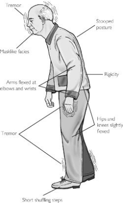
Parkinson’s disease affects several parts of the brain, leading to symptoms like tremor and slow movements.

30-year study

The researchers from Finland’s National Institute for Health and Welfare measured vitamin D levels from the study group between 1978 and 1980, using blood samples.

They then followed these people over 30 years to see whether they developed Parkinson’s disease.

They found that people with the lowest levels of vitamin D were three times more likely to develop Parkinson’s, compared with the group with the highest levels of vitamin D.

Most vitamin D is made by the body when the skin is exposed to sunlight, although some comes from foods like oily fish, milk or cereals.

As people age, however, their skin becomes less able to produce vitamin D.

Doctors have known for many years that vitamin D helps calcium uptake and bone formation.

But research is now showing that it also plays a role in regulating the immune system, as well as in the development of the nervous system.

Vitamin target

Writing in an editorial in the US journal Archives of Neurology, Marian Evatt, assistant professor of neurology at Emory University School of Medicine, says that health authorities should consider raising the target vitamin D level.

“At this point, 30 nanograms per millilitre of blood or more appears optimal for bone health in humans.

“However, researchers don’t yet know what level is optimal for brain health or at what point vitamin D becomes toxic for humans, and this is a topic that deserves close examination.”

Dr Kieran Breen, director of research at Parkinson’s UK, said: “The study provides further clues about the potential environmental factors that may influence or protect against the progression of Parkinson’s.

“A balanced healthy diet should provide the recommended levels of vitamin D.

“Further research is required to find out whether taking a dietary supplement, or increased exposure to sunlight, may have an effect on Parkinson’s, and at what stage these would be most beneficial.”

More on This Story

Related Stories


http://www.bbc.co.uk/news/10601091

—-

Tratamento com a Vitamina D, Cura e Prevenção – Role of Vitamin D in Parkinson’s Disease

parkinsonian-syndrome

VE
US National Library of Medicine
National Institutes of Health

http://www.ncbi.nlm.nih.gov/pmc/

Tratamento com a Vitamina D, Cura e Prevenção – Role of Vitamin D in Parkinson’s Disease

Parte superior do formulário

Role of Vitamin D in Parkinson’s Disease

ISRN Neurol. 2012; 2012: 134289.

Published online 2012 March 7. doi:  10.5402/2012/134289

PMCID: PMC3349248

Role of Vitamin D in Parkinson’s Disease

Khanh Lương * and Lan Nguyễn

Author information ► Article notes ► Copyright and License information ►Go to:


Abstract

Parkinson’s disease (PD) is the second most common form of neurodegeneration in the elderly population. Clinically, it is characterized by tremor, rigidity, slowness of movement, and postural imbalance. A significant association between low serum vitamin D and PD has been demonstrated, suggesting that elevated vitamin D levels might provide protection against PD. Genetic studies have helped identify a number of proteins linking vitamin D to PD pathology, including the major histocompatibility complex (MHC) class II, the vitamin D receptor (VDR), cytochrome P450 2D6 (CYP2D6), chromosome 22, the renin-angiotensin system (RAS), heme oxygenase-1 (HO-1), poly(ADP-ribose) polymerase-1 gene (PARP-1), neurotrophic factor (NTF), and Sp1 transcription factor. Vitamin D has also been implicated in PD through its effects on L-type voltage-sensitive calcium channels (L-VSCC), nerve growth factor (NGF), matrix metalloproteinases (MMPs), prostaglandins (PGs) and cyclooxygenase-2 (COX-2), reactive oxygen species (ROS), and nitric oxide synthase (NOS). A growing body of evidence suggests that vitamin D supplementation may be beneficial for PD patients. Among the different forms of vitamin D, calcitriol (1,25-dihydroxyvitamin D3) is best indicated for PD, because it is a highly active vitamin D3 metabolite with an appropriate receptor in the central nervous system (CNS).


Introduction

Parkinson’s disease (PD) is a movement disorder characterized by tremor, rigidity, slowness of movement, and postural imbalance. There is evidence of abnormalities in the vitamin D-endocrine system in PD patients, including low bone mineral density (BMD), decreased vitamin D levels, and increased bone turnover makers (bone alkaline phosphatase and urinary N-terminal telopeptide of type I collagen) compared to controls [1]. These factors, combined with balance problems, are the probable reasons for the high incidence of fractures, especially of the hip, reported in elderly women with PD [2]. Sunlight exposure can increase the BMD of PD by increasing serum 25-hydroxyvitamin D3 (25OHD) levels [3]. In another study, serum 25OHD and BMD were reported to be reduced in PD patients. The BMD Z-score of the trochanter was directly correlated to the degree of physical activity and total body BMD Z-score correlated to the degree of rigidity [4]. Osteoporosis and osteopenia are common findings in PD patients, affecting up to 91% of women and 61% of men [5]. Decreased BMD, low concentrations of serum ionized calcium, and compensatory hyperparathyroidism increase the risk of hip fracture in PD patients [1]. Despite an abundance of correlational studies, it is unknown whether vitamin D deficiency is a cause or consequence of PD. In the present paper, we review the hypothesized roles of vitamin D in PD pathogenesis.


2. Genomic Factors Associated with Vitamin D in Parkinson’s Disease


2.1. Major Histocompatibility Complex (MHC)

Studies have suggested that several genes in the MHC region promote susceptibility to PD. Significantly increased levels of MHC class II expressions were detected in the cerebrospinal fluid (CSF) monocytes of PD patients [6]. Human leukocyte antigen (HLA) genes have also been implicated in PD, and large numbers of HLA-DR-positive reactive microglia were detected in the substantia nigra (SN) and the nigrostriatal tract in PD patients [78]. HLA-DR-positive microglia have also been found in these regions in a case of Parkinson’s-associated dementia in Guam [8]. Conversely, calcitriol (1,25-dihydroxyvitamin D3) is known to stimulate phagocytosis but suppress MHC class II antigen expression in human mononuclear phagocytes [910]. Calcitriol also decreases interferon-gamma-induced HLA-DR antigen expression on normal and transformed human keratinocytes [1112]. These findings suggested that vitamin D may modulate MHC class II antigen expression in PD.

2.2. Vitamin D Receptor (VDR)

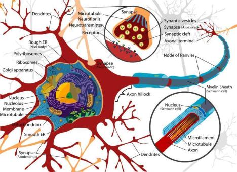
There is ample evidence for vitamin D involvement in mammalian brain function. VDR and 1α-hydroxylase, the enzyme responsible for the formation of active vitamin D in the human brain, are found in the large neurons of the SN, as well as in neurons and glial cells in the hypothalamus [13]. VDR, a nuclear receptor, is restricted to the nucleus but 1α-hydroxylase is distributed throughout the cytoplasm. The presence of these proteins in the CNS suggests that vitamin D is active within the brain. VDR knockout mice have muscular and motor impairment [14]. Genetic studies provide an opportunity to link molecular variations with epidemiological data, DNA sequence variations, such as polymorphisms, exert subtle biological effects on the expression and functionality of proteins. VDR mRNA was identified as a potential blood marker for PD [15]. In a Korean population, the VDR BsmI genotype is reported to be associated with PD [16]. Butler et al. [17] reported that the VDR gene is a potential susceptibility gene for PD in the Caucasian population. These reports suggested a role of vitamin D in PD.

2.3. The Cytochrome P450 (CYP)

CYP superfamily of enzymes is responsible for the oxidation, peroxidation, and/or reduction of vitamins, steroids, and xenobiotics, as well as the metabolism of drugs. CYP2D6, an important member of this superfamily, is expressed in neurons in the brain and gut and may be influenced by polymorphic expression. There is a higher prevalence of the poor-metabolizing CYP2D6*4 allele in PD patients compared with controls (20.7% versus 11.0%) [18]. In case-control and meta-analysis studies, the CYP2D polymorphism was found to be associated with PD [1920]. Other studies, however, did not find an association between the CYP2D6 polymorphism and PD in an Asian population [2122]. Although the poor metabolizer genotype has a small but highly significant association with PD, it would be easily missed in studies with modest numbers of subjects. CYP2D6 protein and enzymatic activity are completely absent in less than 1% of Asian people and in up to 10% of Caucasians with two null alleles [23]. Singh et al. reported the expression of CYP2D22, an ortholog of human CYP2D6, in mouse striatum and its modulation in MPTP-induced PD phenotype and nicotine-mediated neuroprotection [24]. CYP2D6 is a potential 25-hydroxylase, which converts vitamin D3 into 25OHD, and vitamin D 25-hydroxylase deficiency resulted in vitamin D deficiency [25]. Moreover, the CYP2D and PD loci are located on the same chromosome 22 [2627]. Deletion of chromosome 22q11 syndrome was reported to be associated with PD [2829]. Interestingly, patients with a deletion of chromosome 22q11 showed a reduced BMD, serum calcium, and PTH levels; 11% and 8% of these patients had serum 25OHD levels under 20ng/ml and abnormal serum 1,25OHD levels, respectively [30].


2.4. The Renin-Angiotensin System (RAS)

The primary function of the RAS is to maintain fluid homeostasis and regulate blood pressure. Several components of the RAS and its receptors are found in the CNS [3134], suggesting that RAS is important in the brain. CSF levels of angiotensin-converting enzyme (ACE) activity were reported to be decreased in PD patients and increased with dopaminergic treatment [3536]. In addition, the ACE inhibitor perindopril has been shown to exert beneficial effects on the dopaminergic system [3738]. After four weeks of treatment with perindopril, patients with PD had faster improvement in motor response after L-dopa and a reduction in “on phase” peak dyskinesia [39]. The frequency of the homozygous DD genotype of the ACE gene was significantly increased in patients with PD, and is also higher in PD patients with L-dopa-induced psychosis versus without psychosis [4041]. However, other studies did not reveal any associations between ACE polymorphisms, PD, and of L-dopa-induced adverse effects [4243]. The dissimilar findings may be attributable to differences between Chinese and Caucasian populations. Interestingly, there is also an interaction between vitamin D and the RAS. The use of ACE inhibitors by the ACE DD genotype has been shown to decrease the level of calcitriol [44]. In addition, genetic disruption of the VDR resulted in overstimulation of the RAS with increased production of renin and angiotensin II, thereby leading to high blood pressure and cardiac hypertrophy. Treatment with captopril reduced cardiac hypertrophy in VDR knockout mice [45], suggesting that calcitriol may function as an endocrine suppressor of renin biosynthesis. Vitamin D has also been reported to decrease ACE activity in bovine endothelial cells [46]. The findings suggested that vitamin D might affect ACE activity in PD.

2.5. Heme Oxygenase-1 (HO-1)

HO-1 is a stress protein that may confer cytoprotection by enhancing catabolism of pro-oxidant heme to the radical scavenging bile pigments, biliverdin, and bilirubin. The HO-1 gene can be upregulated by a host of noxious stimuli and is induced in CNS tissues affected by neurological diseases [47]. In the normal brain, basal HO-1 expression is low and restricted to small groups of scattered neurons and neuroglia [48]. In the brains of PD patients, the HO-1 is highly overexpressed in astrocytes within the SN and in Lewy bodies found in affected dopaminergic neurons [49]. Serum HO-1 levels are increased in PD patients but not in patients with Alzheimer’s disease (AD) [50], suggesting a systemic antioxidant reaction to a chronic oxidative stress state that is unique to PD. Similarly, calcitriol delayed of HO-1 immunoreactivity after the postlesional survival time of 12 hours concomitant with a reduction in glial fibrillary acidic protein immunoreactivity in remote cortical regions affected by a secondary spread of injury in glial cells of the focal cerebral ischemic [51], thereby supporting the protective role of calcitriol in postcellular injury.

2.6. Poly(ADP-Ribose) Polymerase-1 (PARP-1)

PARP-1 is a nuclear protein that can promote either neuronal death or survival under certain stress conditions. Overexpression of PARP-1 has been reported in the dopaminergic neurons of the SN in PD [52]. PARP-1 is also implicated in 1-methyl-4-phenyl-1,2,3,6-tetrahydropyridine- (MPTP-) induced neurotoxicity in vivo [53]. MPTP is a neurotoxin that induces parkinsonian symptoms in human and animals, but mice lacking PARP gene are spared from MPTP neurotoxicity [54]. Therefore, PARP inhibitors have proved to be valuable tools in PD model [5556]. PARP-1 variants are reported to be protective against PD [57]. In addition, increased levels of vitamin D seem to downregulate PARP-1 expression; PARP-1 levels decrease following calcitriol treatment in NB4 cells, which represent an acute promyelocytic leukemia cells [58]. Vitamin D exerts a concentration-dependent inhibitory effect on PARP-1 in the human keratinocyte cells [59]. Vitamin D-induced downregulation of PARP is further enhanced by nicotinamide in human myeloblastic leukemia cells [60]. Furthermore, PARP is attenuated in the hippocampus of rats that received dexamethasone and vitamin D [61], suggesting that the anti-inflammatory effects of dexamethasone and vitamin D derives from their ability to downregulate microglial activation. These findings suggested that vitamin D may have a protective role in PD by downregulating PARP.

2.7. Neurotrophic Factors (NTFs)

NTFs are keys to surviving various neuronal insults and promote neuronal regeneration following injury [62]. NTFs can exert protective or even restorative effects on PD animal models and dopaminergic cell cultures; key examples include brain-derived neurotrophic factor (BDNF) [63], glial-derived neurotrophic factor (GDNF) [64], mesencephalic-astrocyte-derived neurotrophic factor (MANF) [65], and cerebral dopamine neurotropic factor (CDNF) [66]. Receptors for these NTFs are expressed in the striatum and SN [6768]. NTF expression is reduced in the SN of patients with PD [6971]. Moreover, the C allele of an inotropic CDNF single nucleotide polymorphism (rs7094179) has been suggested to infer susceptibility to PD in a Korean population [72], and A allele of BDNF is associated with PD in a Chinese Han population [73]. Interestingly, calcitriol regulates the expression of the low-affinity neurotrophic receptor [74] and increases striatal GDNF mRNA and protein expression in adult rats [75]. In vivo, calcitriol is also a potent inducer of GDNF in rat glioma cells [76]. The brains of offspring from vitamin D-deficient dams are characterized by diminished expression of neurotrophic factors [77]. Furthermore, calcitriol protects against dopamine loss from 6-hydroxydopamine- (6-OHDA-) lesioned rats by increasing GDNF and partially restores tyrosine hydroxylase expression in SN and striatum [7879].

2.8. Sp1 Transcription Factor


Sp1 transcription factor is a member of an extended family of DNA-binding proteins that is acetylated in response to oxidative stress in neurons [80]. The Sp1 family of proteins plays an important role in controlling the expression of the dopamine transporter gene within dopaminergic neurons [81] and also regulates expression of rat dopamine receptor gene [82]. On the other hand, binding sites for the transcription factor Sp1 have been implicated in the transcription of several genes by hormones. In cultured human fibroblasts, the level of CYP24 (25-OHD 24-hydroxylase) mRNA plays a key role in the metabolism of 1,25OHD and increases up to 20,000-fold in response to calcitriol. Two vitamin D-responsive elements (VDREs) located upstream of the CYP24 gene are primarily responsible for the increased mRNA levels, and Sp1 acted synergistically with these VDREs for the induction [72]. The mVDR promoter is controlled by Sp1 sites [73] and functions as the transactivation component of the VDR/Sp1 complex to trigger gene expression [74]. Moreover, the genes encoding Sp1 and VDR were mapped to human chromosome 12q [7576].


3. Nongenomic Role of Vitamin D in Parkinson’s Disease


3.1. Diabetes Mellitus (DM)

Glucose is the molecule necessary to produce energy in the brain. A link between DM and PD has been suggested in several reports, but the results have been inconsistent. Insulin receptors and their mRNAs are localized in the SN [8384]. A high incidence of abnormal glucose tolerance has been reported in PD and seems to be exacerbated by L-dopa treatment [85]. DM has been associated with the risk of developing PD [8687] whereas others reported from protective to no associations with PD [8890]. Human and experimental animal studies, however, demonstrated neurodegeneration associated with peripheral insulin resistance [91]. In a 6-OHDA model of PD, striatal insulin resistance was observed in the striatum [92], and patients with PD exhibited increased autoimmune reactivity to insulin [93]. Individuals newly diagnosed with PD display reduced insulin-mediated glucose uptake [94], which is hypothesized to be due to inhibit early insulin secretion and hyperglycemia after glucose loading [95]. Furthermore, chronic hyperglycemia decreased limbic extracellular dopamine concentrations and striatal dopaminergic transmission in streptozotocin-induced diabetic rats [9697]. Vitamin D levels may provide a link between the diseases; serum 1,25OHD and 25OHD levels are low in diabetic patients [9899], and diabetic rats had an increased metabolic clearance rate of 1,25OHD [100]. Interestingly, a significant high prevalence of vitamin D insufficiency is reported in patients with PD [101102]. A significant inverse association between serum vitamin D and PD was demonstrated [103] and suggested that high vitamin D status might provide protection against PD. In diabetic-induced rats, vitamin D and insulin treatment markedly recovered the levels of altered gene (cholinergic, dopaminergic, and insulin receptors) expression and its binding parameters nearly to those of the control rats [104]. Maternal vitamin D deficiency was reported to alter the expression of genes involved in dopamine specification in the developing rat mesencephalon [105]. Calcitriol has been shown to protect dopamine neuronal toxicity induced by 6-OHDA and the combination of L-buthionine sulfoximine and MPTP, thereby restoring motor activity in the lesioned animals [106107]. Furthermore, vitamin D was reported to improve rigidity and akinesia and reduce levodopa dosage in a patient with PD [108].

3.2. L-Type Voltage-Sensitive Calcium Channels (L-VSCC)

Unlike most neurons in the brain, dopaminergic neurons function as autonomous pacemakers that rely on L-VSCC to generate action potentials at a clock-like 2–4Hz in the absence of synaptic input [109]. L-VSCC activity during autonomous pacemaking increased the sensitivity of dopaminergic neurons to mitochondrial toxins in an animal model of PD [110]. Epidemiological data supports a link between L-VSCC functioning and the risk of developing PD [111113]. Pretreatment with the calcium channel antagonist nimodipine has been shown to block the development of MPTP-induced neurotoxicity in animal models [114,115]. Israpidine, another L-VSCC antagonist, caused a dose-dependent reduction in L-dopa-induced rotational behavior and abnormal involuntary movements in the 6-OHDA-lesioned rat model of PD [116]. With respect to AD, amyloid-βprotein was reported to promote neurodegeneration by inducing L-VSCC expression and suppressing VDR expression; subsequent treatment with vitamin D protected neurons by preventing cytotoxicity and apoptosis, probably by downregulating L-VSCC and upregulating VDR [117]. Calcitriol decreased L-VSCC activity in aged rats and in neuronal vulnerability with particular impact on reducing age-related changes associated with Ca2+ dysregulation [118119]. Treatment with 24R, 25 dihydroxyvitamin D3 also reduced L-VSCC activity in vascular smooth muscle in rats [120].

3.3. Nerve Growth Factor (NGF)

NGF is a small secreted protein that is important for the growth, maintenance, and survival of certain target neurons. NGF has been implicated in maintaining and regulating the septohippocampal pathway, which is involved in learning and memory [121123]. NGF is also present in the human SN [124] and in the adrenal gland [125]. NGF concentrations are decreased in the SN of the PD and in a rat model of PD [69126]. NGF levels showed greater reductions in early states of the disease compared with advanced stages [126], implying that decreased NGF may be involved in the pathogenesis of PD. NGF is reported to protect dopamine neurotoxicity induced by MPTP, rotenone, and 6-OHDA via different pathways [127129]. The chronic infusion of NGF into the rat striatum resulted in cholinergic hyperinnervation and reduced spontaneous activity of striatal neurons [130]. Moreover, NGF increases survival of dopaminergic grafts, rescues nigral dopaminergic neurons, and restores motor dysfunction in a rat model of PD [131132]. In addition, the brains of newborn rats from vitamin D-deficient dams showed reduced expression of NGF [77]. In vitro, calcitriol regulated the expression of the VDR gene and stimulated the expression of the NGF gene in Schwann cells [133]. In mouse fibroblasts, calcitriol and vitamin D analogs are reported to enhance NGF induction by increasing AP-1 binding activity to the NGF promoter [134135]. These findings suggest a protective role for vitamin D in the CNS.

3.4. Matrix Metalloproteinases (MMPs)

MMPs are proteolytic enzymes responsible for extracellular matrix (ECM) remodeling and the regulation of leukocyte migration through the ECM, which is an important step for inflammatory processes. Neuroinflammation is known to contribute significantly to progressive dopaminergic neurodegeneration in PD. MMP involvement has been reported in the degeneration of dopaminergic neurons. MMP-3 expression is increased during lipopolysaccharide- (LPS-) induced dopamine neurotoxicity [136]. MMP-9 is also elevated in MPTP-induced parkinsonism in mice [137]. Application of dopaminergic neurotoxins to two human neuroblastoma cell lines downregulates the transcription and translation of tissue inhibitor of metalloproteinase- (TIMP-) 2 effectively enhancing MMP activity [138]. Exendin-4, which is an analogue of glucagon-like peptide 1 (GLP-1), significantly attenuates the loss of SN neurons and the striatal dopaminergic fibers in the MPTP-induced PD model, and inhibits the expression of MMP-3 [139]. Conversely, the VDR TaqI polymorphism is associated with decreased production of TIMP-1, a natural inhibitor of MMP-9 [140]. Calcitriol modulates tissue MMP expression under experimental conditions [141]. Calcitriol downregulates MMP-9 levels in keratinocytes and may attenuate deleterious effects due to excessive TNF-α-induced proteolytic activity associated with cutaneous inflammation [142]. In addition, a vitamin D analogue was reported to reduce the expression of MMP-2, MMP-9, VEGF, and parathyroid hormone-related protein in Lewis lung carcinoma cells [143]. These findings suggested that vitamin D plays a role in modulating MMP activation in PD.


3.5. Prostaglandins (PGs)

PGs play a role in inflammatory processes [144]. Cyclooxygenase (COX) participates in the conversion of arachidonic acid into PGs. PGE2 is a key product of COX-2 and is increased in the SN of patients with PD and MPTP-induced PD in an animal model [145146]. PGE2 is also directly and selectively toxic to dopaminergic neurons [147]. PGE2 receptors are found on dopaminergic neurons in the rat SN [147]. Overexpression of COX-2 is reported in PD and an MPTP-animal model [148149]. COX inhibitors provide neuroprotection in the MPTP-mouse model of PD [150]. Similarly, regular use of COX-2 inhibiting nonsteroidal anti-inflammatory drugs (NSAIDs), such as ibuprofen, has been associated with a decreased incidence of PD [151]. Calcitriol, which reportedly regulates the expression of several key genes involved in PG pathways, decreases PG synthesis [152]. Calcitriol and its analogues have also been shown to inhibit selectively the activity of COX-2 [153]. These findings suggested that vitamin D has a role in anti-inflammatory processes in PD.


3.6. Oxidative Stress

Oxidative stress has been suggested to contribute to the pathogenesis of PD. Lymphocytes from untreated PD patients have increase oxidative stress [154]. Analyses of postmortembrains from PD reveal evidence of increased lipid peroxidation in PD SN [155156]. A selective superoxide dismutase (SOD) is also increased in the SN of PD patients [157]. Calcitriol administration has been reported to exert a receptor-mediated effect on the secretion of hydrogen peroxide by human monocytes [158]. In vitro, monocytes gradually lose their ability to produce superoxide when stimulated; the addition of calcitriol, lipopolysaccharide, or lipoteichoic acid (LTA) restored the ability of stimulated monocytes to produce superoxide and increases the oxidative capacity compared with unstimulated monocytes [159]. Calcitriol can also protect nonmalignant prostate cells from oxidative stress-induced cell death by preventing ROS-induced cellular injuries [160]. Vitamin D metabolites and analogues were reported to induce lipoxygenase mRNA expression, lipoxygenase activity, and ROS production in a human bone cell line [161]. Vitamin D can also reduce lipid peroxidation and induce SOD activity in the rat hepatic antioxidant system [162]. These findings suggested a role of vitamin D in oxidative stress in PD.


3.7. Nitric Oxide Synthase (NOS)

NOS is an enzyme involved in the synthesis of nitric oxide (NO), which has also been implicated in PD. In postmortem brains of PD, high levels of NOS expression were found in the nigrostriatal region and basal ganglia [163]. A significant increase in the nitrite content was reported in polymorphonuclear leukocytes of PD patients [164]. Inducible and neuronal NO are increased in both 6-OHDA and MPTP animal models [165166]. Moreover, studies with NOS inhibitors and NOS knock-out animals have also confirmed the role of NOS in neurodegeneration [167168]. Reduced and oxidized glutathione (GSH) were demonstrated in the SN of patients with PD [169]. Conversely, the activation of 1α-hydroxylase in macrophages increases in 1,25OHD, which inhibits inducible NOS (iNOS) expression and reduces NO production by lipopolysaccharide- (LPS-) stimulated macrophages [170]. Thus, calcitriol production by macrophages may provide protection against oxidative injuries caused by the NO burst. Calcitriol is known to inhibit LPS-induced immune activation in human endothelial cells [171]. In experimental allergic encephalomyelitis, calcitriol inhibits the expression of iNOS in the rat CNS [172]. Astrocytes play a pivotal role in CNS detoxification pathways where glutathione (GSH) is involved in the elimination of oxygen and nitrogen reactive species, such as nitric oxide. Calcitriol affects this pathway by enhancing intracellular GSH pools and significantly reduces nitrite production induced by LPS [173].


4. Conclusion

Recent studies have highlighted a possible relationship between vitamin D and PD. Vitamin D may be beneficial in PD patients, as one patient showed improved rigidity and akinesia and was able to decrease their levodopa dosage after vitamin D therapy. Genetic studies have provided opportunities to determine what proteins may link vitamin D to PD pathology. Vitamin D can also act through a number of nongenomic mechanisms, including effects on protein expression, oxidative stress, inflammation, and cellular metabolism. Among the many forms of vitamin D, calcitriol (1,25-dihydroxyvitamin D3) is an attractive therapeutic candidate, because it is a particularly active metabolite, and its receptor is expressed in the CNS.


Conflict of Interests

The authors declare that they have no conflict of interests.


References

1. Abou-Raya S, Helmii M, Abou-Raya A. Bone and mineral metabolism in older adults with Parkinson’s disease. Age and Ageing. 2009;38(6):675–680.[PubMed]

2. Sato Y, Kaji M, Tsuru T, Oizumi K. Risk factors for hip fracture among elderly patients with Parkinson’s disease. Journal of the Neurological Sciences.2001;182(2):89–93. [PubMed]

3. Sato Y, Iwamoto J, Honda Y. Amelioration of osteoporosis and hypovitaminosis D by sunlight exposure in Parkinson’s disease. Parkinsonism and Related Disorders. 2010

4. Lorefält B, Toss G, Granérus A-K. Bone mass in elderly patients with Parkinson’s disease. Acta Neurologica Scandinavica. 2007;116(4):248–254.[PubMed]

5. Invernizzi M, Carda S, Viscontini GS, Cisari C. Osteoporosis in Parkinson’s disease. Parkinsonism and Related Disorders. 2009;15(5):339–346. [PubMed]

6. Fiszer U, Mix E, Fredrikson S, Kostulas V, Link H. Parkinson’s disease and immunological abnormalities: increase of HLA-DR expression on monocytes in cerebrospinal fluid and of CD45RO+ T cells in peripheral blood. Acta Neurologica Scandinavica. 1994;90(3):160–166. [PubMed]

7. McGeer PL, Itagaki S, Boyes BE, McGeer EG. Reactive microglia are positive for HLA-DR in the substantia nigra of Parkinson’s and Alzheimer’s disease brains.Neurology. 1988;38(8):1285–1291. [PubMed]

8. McGeer PL, Itagaki McGeer SEG. Expression of the histocompatibility glycoprotein HLA-DR in neurological disease. Acta Neuropathologica.1988;76(6):550–557. [PubMed]

9. Tokuda N, Levy RB. 1,25-Dihydroxyvitamin D3 stimulates phagocytosis but suppresses HLA-DR and CD13 antigen expression in human mononuclear phagocytes. Proceedings of the Society for Experimental Biology and Medicine.1996;211(3):244–250. [PubMed]

10. Tokuda N, Mizuki N, Kasahara M, Levy RB. 1,25-dihydroxyvitamin D3 down-regulation of HLA-DR on human peripheral blood monocytes. Immunology.1992;75(2):349–354. [PMC free article] [PubMed]

11. Tamaki K, Saitoh A, Kubota Y. 1,25-Dihydroxyvitamin D3 decreases the interferon-γ (IFN-γ) induced HLA-DR expression but not intercellular adhesion molecule 1 (ICAM-1) on human keratinocytes. Regional Immunology.1990;3(5):223–227. [PubMed]

12. Tone T, Eto H, Katsuoka K, Nishioka K, Nishiyama S. Suppression of gamma-interferon induced HLA-DR antigen expression on normal and transformed keratinocytes by 1,25 (OH)2 vitamin D3The Japanese Journal of Dermatology.1991;101(5):519–525. [PubMed]

13. Eyles DW, Smith S, Kinobe R, Hewison M, McGrath JJ. Distribution of the Vitamin D receptor and 1α-hydroxylase in human brain. Journal of Chemical Neuroanatomy. 2005;29(1):21–30. [PubMed]

14. Burne THJ, McGrath JJ, Eyles DW, Mackay-Sim A. Behavioural characterization of Vitamin D receptor knockout mice. Behavioural Brain Research. 2005;157(2):299–308. [PubMed]

15. Scherzer CR, Eklund AC, Morse LJ, et al. Molecular markers of early Parkinson’s disease based on gene expression in blood. Proceedings of the National Academy of Sciences of the United States of America.2007;104(3):955–960. [PMC free article] [PubMed]

16. Kim JS, Kim YI, Song C, et al. Association of vitamin D receptor gene polymorphism and Parkinson’s disease in Koreans. Journal of Korean Medical Science. 2005;20(3):495–498. [PMC free article] [PubMed]

17. Butler MW, Burt A, Edwards TL, et al. Vitamin D receptor gene as a candidate gene for Parkinson disease. Annals of Human Genetics. 2011;75(2):201–210.[PMC free article] [PubMed]

18. Stefanovic M, Topic E, Ivanisevic AM, Relja M, Korsic M. Genotyping of CYP2D6 in Parkinson’s disease. Clinical Chemistry and Laboratory Medicine.2000;38(9):929–934. [PubMed]

19. Singh M, Khanna VK, Shukla R, Parmar D. Association of polymorphism in cytochrome P450 2D6 and N-acetyltransferase-2 with Parkinson’s disease.Disease Markers. 2010;28(2):87–93. [PubMed]

20. McCann SJ, Pond SM, James KM, Le Couteur DG. The association between polymorphisms in the cytochrome P-450 2D6 gene and Parkinson’s disease: a case-control study and meta-analysis. Journal of the Neurological Sciences.1997;153(1):50–53. [PubMed]

21. Ho SL, Kung MHW, Li LSW, Lauder IJ, Ramsden DB. Cytochrome P4502D6 (debrisoquine 4-hydroxylase) and Parkinson’s disease in Chinese and Caucasians.European Journal of Neurology. 1999;6(3):323–329. [PubMed]

22. Woo SI, Kim JW, Seo HG, et al. CYP2D6*4 polymorphism is not associated with Parkinson’s disease and has no protective role against Alzheimer’s disease in the Korean population. Psychiatry and Clinical Neurosciences. 2001;55(4):373–377. [PubMed]

23. Zanger UM, Raimundo S, Eichelbaum M. Cytochrome P450 2D6: overview and update on pharmacology, genetics, biochemistry. Naunyn-Schmiedeberg’s Archives of Pharmacology. 2004;369(1):23–37. [PubMed]

24. Singh S, Singh K, Patel DK, et al. The expression of cyp2d22, an ortholog of human cyp2d6, in mouse striatum and its modulation in 1-methyl 4-phenyl-1,2,3,6-tetrahydropyridine-induced parkinson’s disease phenotype and nicotine-mediated neuroprotection. Rejuvenation Research. 2009;12(3):185–197.[PubMed]

25. Lin CJ, Dardis A, Wijesuriya SD, Abdullah MA, Casella SJ, Miller WL. Lack of mutations in CYP2D6 and CYP27 in patients with apparent deficiency of vitamin D 25-hydroxylase. Molecular Genetics and Metabolism. 2003;80(4):469–472.[PubMed]

26. Shojaee S, Sina F, Banihosseini SS, et al. Genome-wide linkage analysis of a parkinsonian-pyramidal syndrome pedigree by 500 K SNP arrays. American Journal of Human Genetics. 2008;82(6):1375–1384. [PMC free article][PubMed]

27. Wilhelmsen K, Mirel D, Marder K, et al. Is there a genetic susceptibility locus for Parkinson’s disease on chromosome 22q13? Annals of Neurology.1997;41(6):813–817. [PubMed]

28. Zaleski C, Bassett AS, Tam K, Shugar AL, Chow EWC, McPherson E. The Co-occurrence of early onset Parkinson disease and 22q11.2 deletion syndrome.American Journal of Medical Genetics, Part A. 2009;149(3):525–528.[PMC free article] [PubMed]

29. Booij J, Van Amelsvoort T, Boot E. Co-occurrence of early-onset Parkinson disease and 22q11.2 deletion syndrome: potential role for dopamine transporter imaging. American Journal of Medical Genetics A. 2010;152(11):2937–2938.[PubMed]

30. Stagi S, Lapi E, Gambineri E, et al. Bone density and metabolism in subjects with microdeletion of chromosome 22q11 (del22q11) European Journal of Endocrinology. 2010;163(2):329–337. [PubMed]

31. Changaris DG, Keil LC, Severs WB. Angiotensin II immunohistochemistry of the rat brain. Neuroendocrinology. 1978;25(5):257–274. [PubMed]

32. Healy DP, Printz MP. Distribution of immunoreactive angiotensin II, angiotensin I, angiotensinogen, and renin in the central nervous system of intact and nephrectomized rats. Hypertension. 1984;6(supplement 2):130–136.[PubMed]

33. Ganten D, Hermann K, Bayer C. Angiotensin synthesis in the brain and increased turnover in hypertensive rats. Science. 1983;221(4613):869–871.[PubMed]

34. Dzau VJ, Ingelfinger J, Pratt RE, Ellison KE. Identification of renin and angiotensinogen messenger RNA sequences in mouse and rat brains.Hypertension. 1986;8(6):544–548. [PubMed]

35. Zubenko GS, Volicer L, Direnfeld LK. Cerebrospinal fluid levels of angiotensin-converting enzyme in Alzheimer’s disease, Parkinson’s disease and progressive supranuclear palsy. Brain Research. 1985;328(2):215–221. [PubMed]

36. Konings CH, Kuiper MA, Bergmans PLM, Grijpma AM, van Kamp GJ, Wolters CE. Increased angiotensin-eonverting enzyme activity in cerebrospinal fluid of treated patients with Parkinson’s disease. Clinica Chimica Acta. 1994;231(1):101–106. [PubMed]

37. Kurosaki R, Muramatsu Y, Kato H, et al. Effect of angiotensin-converting enzyme inhibitor perindopril on interneurons in MPTP-treated mice. European Neuropsychopharmacology. 2005;15(1):57–67. [PubMed]

38. Jenkins TA, Wong JYF, Howells DW, Mendelsohn FAO, Chai SY. Effect of chronic angiotensin-converting enzyme inhibition on striatal dopamine content in the MPTP-treated mouse. Journal of Neurochemistry. 1999;73(1):214–219.[PubMed]

39. Reardon KA, Mendelsohn FAO, Chai SY, Horne MK. The angiotensin converting enzyme (ACE) inhibitor, perindopril, modifies the clinical features of Parkinson’s disease. Australian and New Zealand Journal of Medicine.2000;30(1):48–53. [PubMed]

40. Lin JJ, Yueh KC, Chang DC, Lin SZ. Association between genetic polymorphism of angiotensin-converting enzyme gene and Parkinson’s disease.Journal of the Neurological Sciences. 2002;199(1-2):25–29. [PubMed]

41. Lin JJ, Yueh KC, Lin SZ, Harn HJ, Liu JT. Genetic polymorphism of the angiotensin converting enzyme and l-dopa-induced adverse effects in Parkinson’s disease. Journal of the Neurological Sciences. 2007;252(2):130–134. [PubMed]

42. Mellick G, Buchanan DD, McCann SJ, et al. The ACE deletion Polymorphism is not associated with Parkinson’s disease. European Neurology. 1999;41(2):103–106. [PubMed]

43. Pascale E, Purcaro C, Passarelli E, et al. Genetic polymorphism of Angiotensin-Converting Enzyme is not associated with the development of Parkinson’s disease and of l-dopa-induced adverse effects. Journal of the Neurological Sciences. 2009;276(1-2):18–21. [PubMed]

44. Pérez-Castrillón JL, Justo I, Sanz A, De Luis D, Dueñas A. Effect of angiotensin converting enzyme inhibitors on 1.25-(OH)2 D levels of hypertensive patients. Relationship with ACE polymorphisms. Hormone and Metabolic Research. 2006;38(12):812–816. [PubMed]

45. Xiang W, Kong J, Chen S, et al. Cardiac hypertrophy in vitamin D receptor knockout mice: role of the systemic and cardiac renin-angiotensin systems.American Journal of Physiology. 2005;288(1):E125–E132. [PubMed]

46. Hagiwara H, Furuhashi H, Nakaya K, Nakamura Y. Effects of vitamin D3 and related compounds on angiotensin converting enzyme activity of endothelial cells and on release of plasminogen activator from them. Chemical and Pharmaceutical Bulletin. 1988;36(12):4858–4864. [PubMed]

47. Hascalovici JR, Vaya J, Khatib S, et al. Brain sterol dysregulation in sporadic AD and MCI: relationship to heme oxygenase-1. Journal of Neurochemistry.2009;110(4):1241–1253. [PMC free article] [PubMed]

48. Barañano DE, Snyder SH. Neural roles for heme oxygenase: contrasts to nitric oxide synthase. Proceedings of the National Academy of Sciences of the United States of America. 2001;98(20):10996–11002. [PMC free article] [PubMed]

49. Castellani R, Smith MA, Richey PL, Perry G. Glycoxidation and oxidative stress in Parkinson disease and diffuse Lewy body disease. Brain Research.1996;737(1-2):195–200. [PubMed]

50. Mateo I, Infante J, Sánchez-Juan P, et al. Serum heme oxygenase-1 levels are increased in Parkinson’s disease but not in Alzheimer’s disease. Acta Neurologica Scandinavica. 2010;121(2):136–138. [PubMed]

51. Oermann E, Bidmon HJ, Witte OW, Zilles K. Effects of 1α,25 dihydroxyvitamin D3 on the expression of HO-1 and GFAP in glial cells of the photothrombotically lesioned cerebral cortex. Journal of Chemical Neuroanatomy. 2004;28(4):225–238. [PubMed]

52. Soós J, Engelhardt JI, Siklós L, Havas L, Majtényi K. The expression of PARP, NF-κB and parvalbumin is increased in Parkinson disease. NeuroReport.2004;15(11):1715–1718. [PubMed]

53. Wang H, Shimoji M, Yu SW, Dawson TM, Dawson VL. Apoptosis inducing factor and PARP-mediated injury in the MPTP mouse model of Parkinson’s disease. Annals of the New York Academy of Sciences. 2003;991:132–139.[PubMed]

54. Mandir AS, Przedborski S, Jackson-Lewis V, et al. Poly(ADP-ribose) polymerase activation mediates 1-methyl-4-phenyl-1,2,3,6-tetrahydropyridine (MPTP)-induced parkinsonism. Proceedings of the National Academy of Sciences of the United States of America. 1999;96(10):5774–5779. [PMC free article][PubMed]

55. Iwashita A, Mihara K, Yamazaki S, et al. A new poly(ADP-ribose) polymerase inhibitor, FR261529 [2-(4-chlorophenyl)-5-quinoxalinecarboxamide], ameliorates methamphetamine-induced dopaminergic neurotoxicity in mice.Journal of Pharmacology and Experimental Therapeutics. 2004;310(3):1114–1124. [PubMed]

56. Yokoyama H, Kuroiwa H, Tsukada T, Uchida H, Kato H, Araki T. Poly(ADP-ribose)polymerase inhibitor can attenuate the neuronal death after 1-methyl-4-phenyl-1,2,3,6-tetrahydropyridine-induced neurotoxicity in mice. Journal of Neuroscience Research. 2010;88(7):1522–1536. [PubMed]

57. Infante J, Sánchez-Juan P, Mateo I, et al. Poly (ADP-ribose) polymerase-1 (PARP-1) genetic variants are protective against Parkinson’s disease. Journal of the Neurological Sciences. 2007;256(1-2):68–70. [PubMed]

58. Bhatia M, Kirkland JB, Mecking-Gill KA. Modulation of poly(ADP-ribose) polymerase during neurophilic and monocytic differentiation of promyelocytic (NB4) and myelocytic (HL-60) leakaemia cells. Biochemical Journal.1995;308:131–137. [PMC free article] [PubMed]

59. Mabley JG, Wallace R, Pacher P, Murphy K, Szabó C. Inhibition of poly(adenosine diphosphate-ribose) polymerase by the active form of vitamin D.International Journal of Molecular Medicine. 2007;19(6):947–952.[PMC free article] [PubMed]

60. Shen M, Yen A. Nicotinamide cooperates with retinoic acid and 1,25-dihydroxyvitamin D3 to regulate cell differentiation and cell cycle arrest of human myeloblastic leukemia cells. Oncology. 2009;76(2):91–100. [PMC free article][PubMed]

61. Moore M, Piazza A, Nolan Y, Lynch MA. Treatment with dexamethasone and vitamin D3 attenuates neuroinflammatory age-related changes in rat hippocampus. Synapse. 2007;61(10):851–861. [PubMed]

62. McAllister AK. Neurotrophins and neuronal differentiation in the central nervous system. Cellular and Molecular Life Sciences. 2001;58(8):1054–1060.[PubMed]

63. Numakawa T, Matsumoto T, Numakawa M, Richards M, Yamawaki S, Kunugi H. Protection action of neurotrophic factors and estrogen against oxidative stress-mediated neurodegeneration. Journal of Toxicology. 2011;2011:12 pages. Article ID 405194. [PMC free article] [PubMed]

64. Garbayo E, Ansorena E, Lanciego JL, Blanco-Prieto MJ, Aymerich MS. Long-term neuroprotection and neurorestoration by glial cell-derived neurotrophic factor microspheres for the treatment of Parkinson’s disease. Movement Disorders. 2011;26(10):1943–1947. [PubMed]

65. Voutilainen MH, Bäck S, Pörsti E, et al. Mesencephalic astrocyte-derived neurotrophic factor is neurorestorative in rat model of Parkinson’s disease.Journal of Neuroscience. 2009;29(30):9651–9659. [PubMed]

66. Sun Z-P, Gong L, Huang S-H, Geng Z, Cheng L, Chen Z-Y. Intracellular trafficking and secretion of cerebral dopamine neurotrophic factor in neurosecretory cells. Journal of Neurochemistry. 2011;117(1):121–132. [PubMed]

67. Yan Q, Radeke MJ, Matheson CR, Talvenheimo J, Welcher AA, Feinstein SC. Immunocytochemical localization of TrkB in the central nervous system of the adult rat. Journal of Comparative Neurology. 1997;378(1):135–157. [PubMed]

68. Marco S, Saura J, Prez-Navarro E, Mart MJ, Tolosa E, Alberch J. Regulation of c-Ret, GFRα1, and GFRα2 in the substantia nigra pars compacta in a rat model of Parkinson’s disease. Journal of Neurobiology. 2002;52(4):343–351. [PubMed]

69. Mogi M, Togari A, Kondo T, et al. Brain-derived growth factor and nerve growth factor concentrations are decreased in the substantia nigra in Parkinson’s disease. Neuroscience Letters. 1999;270(1):45–48. [PubMed]

70. Howells DW, Porritt MJ, Wong JYF, et al. Reduced BDNF mRNA expression in the Parkinson’s disease substantia nigra. Experimental Neurology.2000;166(1):127–135. [PubMed]

71. Scalzo P, Kümmer A, Bretas TL, Cardoso F, Teixeira AL. Serum levels of brain-derived neurotrophic factor correlate with motor impairment in Parkinson’s disease. Journal of Neurology. 2010;257(4):540–545. [PubMed]

72. Choi J-M, Hong J-H, Chae M-J, et al. Analysis of mutations and the association between polymorphisms in the cerebral dopamine neurotrophic factor (CDNF) gene and Parkinson disease. Neuroscience Letters. 2011;493(3):97–101.[PubMed]

73. Chen L, Wang Y, Xiao H, et al. The 712A/G polymorphism of Brain-derived neurotrophic factor is associated with Parkinson’s disease but not Major Depressive Disorder in a Chinese Han population. Biochemical and Biophysical Research Communications. 2011;408(2):318–321. [PubMed]

74. Naveilhan P, Neveu I, Baudet C, et al. 1,25-dihydroxyvitamin D3 regulates the expression of the low-affinity neurotrophin receptor. Molecular Brain Research.1996;41(1-2):259–268. [PubMed]

75. Sanchez B, Lopez-Martin E, Segura C, Labandeira-Garcia JL, Perez-Fernandez R. 1,25-Dihydroxyvitamin D3 increases striatal GDNF mRNA and protein expression in adult rats. Molecular Brain Research. 2002;108(1-2):143–146.[PubMed]

76. Naveilhan P, Neveu I, Wion D, Brachet P. 1,25-Dihydroxyvitamin D3, an inducer of glial cell line-derived neurotrophic factor. NeuroReport.1996;7(13):2171–2175. [PubMed]

77. Féron F, Burne THJ, Brown J, et al. Developmental Vitamin D3 deficiency alters the adult rat brain. Brain Research Bulletin. 2005;65(2):141–148.[PubMed]

78. Smith MP, Fletcher-Turner A, Yurek DM, Cass WA. Calcitriol protection against dopamine loss induced by intracerebroventricular administration of 6-hydroxydopamine. Neurochemical Research. 2006;31(4):533–539. [PubMed]

79. Sanchez B, Relova JL, Gallego R, Ben-Batalla I, Perez-Fernandez R. 1,25-Dihydroxyvitamin D3 administration to 6-hydroxydopamine-lesioned rats increases glial cell line-derived neurotrophic factor and partially restores tyrosine hydroxylase expression in substantia nigra and striatum. Journal of Neuroscience Research. 2009;87(3):723–732. [PubMed]

80. Ryu H, Lee J, Zaman K, et al. Sp1 and Sp3 are oxidative stress-inducible, antideath transcription factors in cortical neurons. Journal of Neuroscience.2003;23(9):3597–3606. [PubMed]

81. Wang J, Bannon MJ. Sp1 and Sp3 activate transcription of the human dopamine transporter gene. Journal of Neurochemistry. 2005;93(2):474–482.[PubMed]

82. Yajima S, Lee SH, Minowa T, Mouradian MM. Sp family transcription factors regulate expression of rat D2 dopamine receptor gene. DNA and Cell Biology.1998;17(5):471–479. [PubMed]

83. Figlewicz DP, Evans SB, Murphy J, Hoen M, Baskin DG. Expression of receptors for insulin and leptin in the ventral tegmental area/substantia nigra (VTA/SN) of the rat. Brain Research. 2003;964(1):107–115. [PubMed]

84. Takahashi M, Yamada T, Tooyama I, et al. Insulin receptor mRNA in the substantia nigra in Parkinson’s disease. Neuroscience Letters. 1996;204(3):201–204. [PubMed]

85. Sandyk R. The relationship between diabetes mellitus and Parkinson’s disease. International Journal of Neuroscience. 1993;69(1–4):125–130.[PubMed]

86. D’Amelio M, Ragonese P, Callari G, et al. Diabetes preceding Parkinson’s disease onset. A case-control study. Parkinsonism and Related Disorders.2009;15(9):660–664. [PubMed]

87. Xu Q, Park Y, Huang X, et al. Diabetes and risk of Parkinson’s disease.Diabetes Care. 2011;34(4):910–915. [PMC free article] [PubMed]

88. Schernhammer E, Hansen J, Rugbjerg K, Wermuth L, Ritz B. Diabetes and the risk of developing Parkinson’s disease in Denmark. Diabetes Care.2011;34(5):1102–1108. [PMC free article] [PubMed]

89. Powers KM, Smith-Weller T, Franklin GM, Longstreth WT, Swanson PD, Checkoway H. Diabetes, smoking, and other medical conditions in relation to Parkinson’s disease risk. Parkinsonism and Related Disorders. 2006;12(3):185–189. [PubMed]

90. Becker C, Brobert GP, Johansson S, Jick SS, Meier CR. Diabetes in patients with idiopathic parkinson’s disease. Diabetes Care. 2008;31(9):1808–1812.[PMC free article] [PubMed]

91. Fishel MA, Watson GS, Montine TJ, et al. Hyperinsulinemia provokes synchronous increases in central inflammation and β-amyloid in normal adults.Archives of Neurology. 2005;62(10):1539–1544. [PubMed]

92. Morris JK, Zhang H, Gupte AA, Bomhoff GL, Stanford JA, Geiger PC. Measures of striatal insulin resistance in a 6-hydroxydopamine model of Parkinson’s disease. Brain Research. 2008;1240(C):185–195. [PMC free article][PubMed]

93. Wilhelm KR, Yanamandra K, Gruden MA, et al. Immune reactivity towards insulin, its amyloid and protein S100B in blood sera of Parkinson’s disease patients. European Journal of Neurology. 2007;14(3):327–334. [PubMed]

94. Van Woert MH, Mueller PS. Glucose, insulin, and free fatty acid metabolism in Parkinson’s disease treated with levodopa. Clinical Pharmacology and Therapeutics. 1971;12(2):360–367. [PubMed]

95. Boyd 3rd. A.E. AE, Lebovitz HE, Feldman JM. Endocrine function and glucose metabolism in patients with Parkinson’s disease and their alternation by L-Dopa.Journal of Clinical Endocrinology and Metabolism. 1971;33(5):829–837.[PubMed]

96. Murzi E, Contreras Q, Teneud L, et al. Diabetes decreases limbic extracellular dopamine in rats. Neuroscience Letters. 1996;202(3):141–144. [PubMed]

97. Shimizu H, Shimomura Y, Takahashi M, Kobayashi I, Kobayashi S. Dopamine receptor in the streptozotocin-induced diabetic rats. Experimental and Clinical Endocrinology. 1990;95(2):263–266. [PubMed]

98. Kaur H, Donaghue KC, Chan AK, et al. Vitamin D deficiency is associated with retinopathy in children and adolescents with type 1 diabetes. Diabetes Care.2011;34(6):1400–1402. [PMC free article] [PubMed]

99. Yiu Y-F, Chan Y-H, Yiu K-H, et al. Vitamin D deficiency is associated with depletion of circulating endothelial progenitor cells and endothelial dysfunction in patients with type 2 diabetes. Journal of Clinical Endocrinology and Metabolism. 2011;96(5):E830–E835. [PubMed]

100. Verhaeghe J, Suiker AMH, Van Bree R, et al. Increased clearance of 1,25(OH)2D3 and tissue-specific responsiveness to 1,25(OH)2D3 in diabetic rats.American Journal of Physiology. 1993;265(2):E215–E223. [PubMed]

101. Sato Y, Kikuyama M, Oizumi K. High prevalence of vitamin D deficiency and reduced bone mass in Parkinson’s disease. Neurology. 1997;49(5):1273–1278.[PubMed]

102. Evatt ML, DeLong MR, Kumari M, Auinger P, McDermott MP, Tangpricha V. High prevalence of hypovitaminosis D status in patients with early Parkinson disease. Archives of Neurology. 2011;68(3):314–319. [PubMed]

103. Knekt P, Kilkkinen A, Rissanen H, Marniemi J, Sääksjärvi K, Heliövaara M. Serum vitamin D and the risk of Parkinson disease. Archives of Neurology.2010;67(7):808–811. [PMC free article] [PubMed]

104. Peeyush KT, Savitha B, Sherin A, Anju TR, Jes P, Paulose CS. Cholinergic, dopaminergic and insulin receptors gene expression in the cerebellum of streptozotocin-induced diabetic rats: functional regulation with Vitamin D3supplementation. Pharmacology Biochemistry and Behavior. 2010;95(2):216–222. [PubMed]

105. Cui X, Pelekanos M, Burne THJ, McGrath JJ, Eyles DW. Maternal vitamin D deficiency alters the expression of genes involved in dopamine specification in the developing rat mesencephalon. Neuroscience Letters. 2010;486(3):220–223.[PubMed]

106. Wang JY, Wu JN, Cherng TL, et al. Vitamin D3 attenuates 6-hydroxydopamine-induced neurotoxicity in rats. Brain Research.2001;904(1):67–75. [PubMed]

107. Shinpo K, Kikuchi S, Sasaki H, Moriwaka F, Tashiro K. Effect of 1,25-dihydroxyvitamin D3 on cultured mesencephalic dopaminergic neurons to the combined toxicity caused by L-buthionine sulfoximine and 1-methyl-4-phenylpyridine. Journal of Neuroscience Research. 2000;62(3):374–382.[PubMed]

108. Derex L, Trouillas P. Reversible Parkinsonism, hypophosphoremia, and hypocalcemia under vitamin D therapy: case report. Movement Disorders.1997;12(4):612–613. [PubMed]

109. Surmeier DJ. Calcium, ageing, and neuronal vulnerability in Parkinson’s disease. Lancet Neurology. 2007;6(10):933–938. [PubMed]

110. Martella G, Madeo G, Schirinzi T, et al. Altered profile and D2-dopamine receptor modulation of high voltage-activated calcium current in striatal medium spiny neurons from animal models of Parkinson’s disease. Neuroscience.2011;177:240–251. [PubMed]

111. Lee CR, Tepper JM. A calcium-activated nonselective cation conductance underlies the plateau potential in rat substantia nigra GABAergic neurons.Journal of Neuroscience. 2007;27(24):6531–6541. [PubMed]

112. Chan CS, Gertler TS, Surmeier DJ. A molecular basis for the increased vulnerability of substantia nigra dopamine neurons in aging and Parkinson’s disease. Movement Disorders. 2010;25(1):S63–S70. [PubMed]

113. Chan CS, Gertler TS, Surmeier DJ. Calcium homeostasis, selective vulnerability and Parkinson’s disease. Trends in Neurosciences. 2009;32(5):249–256. [PubMed]

114. Kupsch A, Gerlach M, Pupeter SC, et al. Pretreatment with nimodipine prevents MPTP-induced neurotoxicity at the nigral, but not at the striatal level in mice. NeuroReport. 1995;6(4):621–625. [PubMed]

115. Kupsch A, Sautter J, Schwarz J, Riederer P, Gerlach M, Oertel WH. 1-methyl-4-phenyl-1,2,3,6-tetrahydropyridine-induced neurotoxicity in non-human primates is antagonized by pretreatment with nimodipine at the nigral, but not at the striatal level. Brain Research. 1996;741(1-2):185–196. [PubMed]

116. Schuster S, Doudnikoff E, Rylander D, et al. Antagonizing L-type Ca2+Channel Reduces Development of Abnormal Involuntary Movement in the Rat Model of L-3,4-Dihydroxyphenylalanine-Induced Dyskinesia. Biological Psychiatry. 2009;65(6):518–526. [PubMed]

117. Dursun E, Gezen-Ak D, Yilmazer S. A novel perspective for Alzheimer’s disease: vitamin D receptor suppression by amyloid-β and preventing the amyloid-β induced alterations by vitamin D in cortical neurons. Journal of Alzheimer’s Disease. 2011;23(2):207–219. [PubMed]

118. Brewer LD, Porter NM, Kerr DS, Landfield PW, Thibault O. Chronic 1α,25-(OH)2vitamin D3 treatment reduces Ca2+-mediated hippocampal biomarkers of aging. Cell Calcium. 2006;40(3):277–286. [PubMed]

119. Brewer LD, Thibault V, Chen KC, Langub MC, Landfield PW, Porter NM. Vitamin D hormone confers neuroprotection in parallel with downregulation of L-type calcium channel expression in hippocampal neurons. Journal of Neuroscience. 2001;21(1):98–108. [PubMed]

120. Shan JJ, Li B, Taniguchi N, Pang PKT. Inhibition of membrane L-type calcium channel activity and intracellular calcium concentration by 24R,25-dihydroxyvitamin D3 in vascular smooth muscle. Steroids. 1996;61(11):657–663.[PubMed]

121. Wu CWK, Yeh HH. Nerve growth factor rapidly increases muscarinic tone in mouse medial septum/diagonal band of Broca. Journal of Neuroscience.2005;25(17):4232–4242. [PubMed]

122. Poucet B, Herrmann T. Septum and medial frontal cortex contribution to spatial problem-solving. Behavioural Brain Research. 1990;37(3):269–280.[PubMed]

123. Olton DS, Walker JA, Gage FH. Hippocampal connections and spatial discrimination. Brain Research. 1978;139(2):295–308. [PubMed]

124. Nishio T, Furukawa S, Akiguchi I, Sunohara N. Medial nigral dopamine neurons have rich neurotrophin support in humans. NeuroReport.1998;9(12):2847–2851. [PubMed]

125. Silani V, Pizzuti A, Falini A, et al. β-Nerve growth factor (β-NGF) mRNA expression in the Parkinsonian adrenal gland. Experimental Neurology.1991;113(2):166–170. [PubMed]

126. Lorigados Pedre L, Pavón Fuentes N, Alvarez González L, et al. Nerve growth factor levels in parkinson disease and experimental parkinsonian rats. Brain Research. 2002;952(1):122–127. [PubMed]

127. Shimoke K, Chiba H. Nerve growth factor prevents 1-methyl-4-phenyl-1,2,3,6-tetrahydropyridine-induced cell death via the Akt pathway by suppressing caspase-3-like activity using PC12 cells: relevance to therapeutical application for Parkinson’s disease. Journal of Neuroscience Research. 2001;63(5):402–409.[PubMed]

128. Hirata Y, Meguro T, Kiuchi K. Differential effect of nerve growth factor on dopaminergic neurotoxin-induced apoptosis. Journal of Neurochemistry.2006;99(2):416–425. [PubMed]

129. Salinas M, Diaz R, Abraham NG, De Galarreta CMR, Cuadrado A. Nerve growth factor protects against 6-hydroxydopamine-induced oxidative stress by increasing expression of heme oxygenase-1 in a phosphatidylinositol 3-kinase-dependent manner. Journal of Biological Chemistry. 2003;278(16):13898–13904. [PubMed]

130. Forander P, Soderstrom S, Humpel C, Strömberg I. Chronic infusion of nerve growth factor into rat striatum increases cholinergic markers and inhibits striatal neuronal discharge rate. European Journal of Neuroscience. 1996;8(9):1822–1832. [PubMed]

131. Chaturvedi RK, Shukla S, Seth K, Agrawal AK. Nerve growth factor increases survival of dopaminergic graft, rescue nigral dopaminergic neurons and restores functional deficits in rat model of Parkinson’s disease. Neuroscience Letters.2006;398(1-2):44–49. [PubMed]

132. Li M, Zhang SZ, Guo YW, et al. Human umbilical vein-derived dopaminergic-like cell transplantation with nerve growth factor ameliorates motor dysfunction in a rat model of parkinson’s disease. Neurochemical Research. 2010;35(10):1522–1529. [PubMed]

133. Cornet A, Baudet C, Neveu I, Baron-Van Evercooren A, Brachet P, Naveilhan P. 1,25-Dihydroxyvitamin D3 regulates the expression of VDR and NGF gene in Schwann cells in vitro. Journal of Neuroscience Research. 1998;53(6):742–746.[PubMed]

134. Musiol IM, Feldman D. 1,25-dihydroxyvitamin D3 induction of nerve growth factor in L929 mouse fibroblasts: effect of vitamin D receptor regulation and potency of vitamin D3 analogs. Endocrinology. 1997;138(1):12–18. [PubMed]

135. Veenstra TD, Fahnestock M, Kumar R. An AP-1 site in the nerve growth factor promoter is essential for 1,25-dihydroxyvitamin D3-mediated nerve growth factor expression in osteoblasts. Biochemistry. 1998;37(17):5988–5994.[PubMed]

136. McClain JA, Phillips LL, Fillmore HL. Increased MMP-3 and CTGF expression during lipopolysaccharide-induced dopaminergic neurodegeneration.Neuroscience Letters. 2009;460(1):27–31. [PubMed]

137. Lorenzl S, Calingasan N, Yang L, et al. Matrix metalloproteinase-9 is elevated in 1-methyl-4-phenyl-1,2,3,6-tetrahydropyridine-induced parkinsonism in mice. NeuroMolecular Medicine. 2004;5(2):119–131. [PubMed]

138. Kim SY, Woo MS, Park JS, Hyun JW, Kim YS, Kim HS. The neuroprotective role of tissue inhibitor of metalloproteinase-2 in MPP+– or 6-OHDA-treated SK-N-BE(2)C and SH-SY5Y human neuroblastoma cells. Neuroscience Letters.2010;468(2):136–140. [PubMed]

139. Kim S, Moon M, Park S. Exendin-4 protects dopaminergic neurons by inhibition of microglial activation and matrix metalloproteinase-3 expression in an animal model of Parkinson’s disease. Journal of Endocrinology.2009;202(3):431–439. [PubMed]

140. Timms PM, Mannan N, Hitman GA, et al. Circulating MMP9, vitamin D and variation in the TIMP-1 response with VDR genotype: mechanisms for inflammatory damage in chronic disorders? Monthly Journal of the Association of Physicians. 2002;95(12):787–796. [PubMed]

141. Dean DD, Schwartz Z, Schmitz J, et al. Vitamin D regulation of metalloproteinase activity in matrix vesicles. Connective Tissue Research.1996;35(1–4):331–336. [PubMed]

142. Bahar-Shany K, Ravid A, Koren R. Upregulation of MMP-9 production by TNFα in keratinocytes and its attenuation by vitamin D. Journal of Cellular Physiology. 2010;222(3):729–737. [PubMed]

143. Nakagawa K, Sasaki Y, Kato S, Kubodera N, Okano T. 22-Oxa-1α,25-dihydroxyvitamin D3 inhibits metastasis and angiogenesis in lung cancer.Carcinogenesis. 2005;26(6):1044–1054. [PubMed]

144. Ricciotti E, Fitzgerald GA. Prostaglandins and inflammation.Arteriosclerosis, Thrombosis, and Vascular Biology. 2011;31(5):986–1000.[PMC free article] [PubMed]

145. Mattammal MB, Strong R, Lakshmi VM, Chung HD, Stephenson AH. Prostaglandin H synthetase-mediated metabolism of dopamine: implication for Parkinson’s disease. Journal of Neurochemistry. 1995;64(4):1645–1654.[PubMed]

146. Wang T, Pei Z, Zhang W, et al. MPP+-induced COX-2 activation and subsequent dopaminergic neurodegeneration. FASEB Journal. 2005;19(9):1134–1136. [PubMed]

147. Carrasco E, Casper D, Werner P. PGE2 receptor EP1 renders dopaminergic neurons selectively vulnerable to low-level oxidative stress and direct PGE2neurotoxicity. Journal of Neuroscience Research. 2007;85(14):3109–3117.[PubMed]

148. Knott C, Stern G, Wilkin GP. Inflammatory regulators in Parkinson’s disease: iNOS, lipocortin-1, and cyclooxygenases-1 and -2. Molecular and Cellular Neuroscience. 2000;16(6):724–739. [PubMed]

149. Teismann P, Tieu K, Choi DK, et al. Cyclooxygenase-2 is instrumental in Parkinson’s disease neurodegeneration. Proceedings of the National Academy of Sciences of the United States of America. 2003;100(9):5473–5478.[PMC free article] [PubMed]

150. Teismann P, Ferger B. Inhibition of the cyclooxygenase isoenzymes COX-1 and COX-2 provide neuroprotection in the MPTP-mouse model of Parkinson’s disease. Synapse. 2001;39(2):167–174. [PubMed]

151. Chen H, Zhang SM, Hernán MA, et al. Nonsteroidal anti-inflammatory drugs and the risk of Parkinson disease. Archives of Neurology. 2003;60(8):1059–1064. [PubMed]

152. Moreno J, Krishnan AV, Swami S, Nonn L, Peehl DM, Feldman D. Regulation of prostaglandin metabolism by calcitriol attenuates growth stimulation in prostate cancer cells. Cancer Research. 2005;65(17):7917–7925.[PubMed]

153. Aparna R, Subhashini J, Roy KR, et al. Selective inhibition of cyclooxygenase-2 (COX-2) by 1 α,25-dihydroxy- 16-ene-23-yne-vitamin D3, a less calcemic vitamin D analog. Journal of Cellular Biochemistry. 2008;104(5):1832–1842. [PubMed]

154. Prigione A, Isaias IU, Galbussera A, et al. Increased oxidative stress in lymphocytes from untreated Parkinson’s disease patients. Parkinsonism and Related Disorders. 2009;15(4):327–328. [PubMed]

155. Dexter DT, Carter CJ, Wells FR, et al. Basal lipid peroxidation in substantia nigra is increased in Parkinson’s disease. Journal of Neurochemistry.1989;52(2):381–389. [PubMed]

156. Dexter DT, Holley AE, Flitter WD, et al. Increased levels of lipid hydroperoxides in the Parkinsonian Substantia nigra: an HPLC and ESR study.Movement Disorders. 1994;9(1):92–97. [PubMed]

157. Saggu H, Cooksey J, Dexter D, et al. A selective increase in particulate superoxide dismutase activity in parkinsonian substantia nigra. Journal of Neurochemistry. 1989;53(3):692–697. [PubMed]

158. Tesco G, Latorraca S, Piersanti P, Sorbi S, Piacentini S, Amaducci L. Free radical injury in skin cultured fibroblasts from Alzheimer’s disease patients.Annals of the New York Academy of Sciences. 1992;673:149–153. [PubMed]

159. Cohen MS, Mesler DE, Snipes RG, Gray TK. 1,25-Dihydroxyvitamin D3activates secretion of hydrogen peroxide by human monocytes. Journal of Immunology. 1986;136(3):1049–1053. [PubMed]

160. Levy R, Malech HL. Effect of 1,25-dihydroxyvitamin D3, lipopolysaccharide, or lipoteichoic acid on the expression of NADPH oxidase components in cultured human monocytes. Journal of Immunology. 1991;147(9):3066–3071. [PubMed]

161. Bao BY, Ting HJ, Hsu JW, Lee YF. Protective role of 1α, 25-dihydroxyvitamin D3 against oxidative stress in nonmalignant human prostate epithelial cells.International Journal of Cancer. 2008;122(12):2699–2706. [PubMed]

162. Somjen D, Katzburg S, Grafi-Cohen M, Knoll E, Sharon O, Posner GH. Vitamin D metabolites and analogs induce lipoxygenase mRNA expression and activity as well as reactive oxygen species (ROS) production in human bone cell line. Journal of Steroid Biochemistry and Molecular Biology. 2011;123(1-2):85–89. [PubMed]

163. Hunot S, Boissière F, Faucheux B, et al. Nitric oxide synthase and neuronal vulnerability in Parkinson’s disease. Neuroscience. 1996;72(2):355–363.[PubMed]

164. Barthwal MK, Srivastava N, Shukla R, et al. Polymorphonuclear leukocyte nitrite content and antioxidant enzymes in Parkinson’s disease patients. Acta Neurologica Scandinavica. 1999;100(5):300–304. [PubMed]

165. Singh S, Das T, Ravindran A, et al. Involvement of nitric oxide in neurodegeneration: a study on the experimental models of Parkinson’s disease.Redox Report. 2005;10(2):103–109. [PubMed]

166. Muramatsu Y, Kurosaki R, Watanabe H, et al. Cerebral alterations in a MPTP-mouse model of Parkinson’s disease—an immunocytochemical study.Journal of Neural Transmission. 2003;110(10):1129–1144. [PubMed]

167. Schulz JB, Matthews RT, Muqit MMK, Browne SE, Beal MF. Inhibition of neuronal nitric oxide synthase by 7-nitroindazole protects against MPTP-induced neurotoxicity in mice. Journal of Neurochemistry. 1995;64(2):936–939.[PubMed]

168. Dehmer T, Lindenau J, Haid S, Dichgans J, Schulz JB. Deficiency of inducible nitric oxide synthase protects against MPTP toxicity in vivo. Journal of Neurochemistry. 2000;74(5):2213–2216. [PubMed]

169. Kaur D, Lee D, Ragapolan S, Andersen JK. Glutathione depletion in immortalized midbrain-derived dopaminergic neurons results in increases in the labile iron pool: implications for Parkinson’s disease. Free Radical Biology and Medicine. 2009;46(5):593–598. [PMC free article] [PubMed]

170. Chang JM, Kuo MC, Kuo HT, et al. 1-α,25-Dihydroxyvitamin D3 regulates inducible nitric oxide synthase messenger RNA expression and nitric oxide release in macrophage-like RAW 264.7 cells. Journal of Laboratory and Clinical Medicine. 2004;143(1):14–22. [PubMed]

171. Garcion E, Nataf S, Berod A, Darcy F, Brachet P. 1,25-Dihydroxyvitamin D3inhibits the expression of inducible nitric oxide synthase in rat central nervous system during experimental allergic encephalomyelitis. Molecular Brain Research. 1997;45(2):255–267. [PubMed]

172. Equils O, Naiki Y, Shapiro AM, et al. 1,25-Dihydroxyvitamin D3 inhibits lipopolysaccharide-induced immune activation in human endothelial cells.Clinical and Experimental Immunology. 2006;143(1):58–64. [PMC free article][PubMed]

173. Garcion E, Sindji L, Leblondel G, Brachet P, Darcy F. 1,25-dihydroxyvitamin D3 regulates the synthesis of γ-glutamyl transpeptidase and glutathione levels in rat primary astrocytes. Journal of Neurochemistry. 1999;73(2):859–866.[PubMed]


Articles from ISRN Neurology are provided here courtesy of Hindawi Publishing Corporation


Formats:

|

  • Article

|

|

|


Related citations in PubMed

See reviews…See all…

Links

Recent activity

ClearTurn Off

PMC

PubMed

See more…

See more …

See more …

See more …

See more …

See more …

You are here: NCBI > Literature > PubMed Central (PMC)

—-

Tratamento com a Vitamina D, Cura e Prevenção – Parkinsonian Syndrome and Essential Tremor Movement Disorders

Tratamento com a Vitamina D, Cura e Prevenção – Parkinsonian Syndrome and Essential Tremor Movement

ACOMPANHE AS PUBLICAÇOES SOBRE Tratamento com a Vitamina D, Cura e Prevenção

VE


If you have a parkinsonian syndrome (PS) or essential tremor (ET)movement disorder, you may have trouble with certain forms of movement, from turning over in bed, to doing buttons, to swinging your arms as you walk. Controlling everyday actions can be a struggle.

A diagnosis of a PS or ET movement disorder doesn’t mean that you can’t be physically active; in fact, many researchers agree that regular exercise, such as walking, hiking, or dancing, is good for people with movement disorders. Moving can help rebuild strength and coordination. Talk to your doctor before you get started to figure out what type and how much exercise you can do.

Next steps

If you suspect that you have a PS or ET movement disorder, it’s important to get the right diagnosis. Start now by learning about these different types of movement disorders and how the brain is involved.

The Brain’s Role in Parkinsonian Syndrome and Essential Tremor

Read about how PS and ET movement disorders begin in the brain.

The Differences Between Parkinsonian Syndrome and Essential Tremor

Learn the similarities and differences between PS and ET movement disorders.

http://move2answers.gehealthcare.com/movement-disorders/

—-

Link Between Vitamin D and Parkinson’s Disease

Low Vitamin D Levels Seen in Parkinson’s Patients  


Study Suggests Possible Link Between Vitamin D and Parkinson’s Disease


parc By Salynn Boyles WebMD Health News Reviewed by Laura J. Martin, MD


March 14, 2011 — A study of newly diagnosed patients with Parkinson’s disease found a high prevalence of vitamin Dinsufficiency, but vitamin D levels did not continue to decline as the disease progressed.


The research is one of several studies suggesting a link between low vitamin D levels and Parkinson’s disease, a brain disorder that leads to tremors and problems with balance and coordination. Parkinson’s affects as many as 1 million older Americans.


In a study from Finland published last summer, people with the lowest levels of vitamin D were significantly more likely to develop Parkinson’s over almost three decades of follow-up, compared to people with the highest blood levels of the vitamin. In the newly published study, almost 70% of patients with a recent diagnosis of Parkinson’s disease had low blood levels of the vitamin. But it is not yet clear if vitamin D insufficiency raises Parkinson’s risk or if having high levels of the vitamin is protective, says study researcher Marian L. Evatt, MD, of Emory University School of Medicine and the Atlanta Veterans Affairs Medical Center.


“More research is needed to figure this out,” she tells WebMD. “There is certainly an association, but we can’t say if it is causal.”


Does Vitamin D Protect the Brain?


Most people get the majority of their vitamin D from exposure to sunlight. Salmon, tuna, and fortified milk and other dairy products are the main food sources of the vitamin. There are suggestions from animal and other studies that vitamin D protects the brain and central nervous system.


If this is the case, it would stand to reason that people with low vitamin D levels would have an increased risk for developing Parkinsons’ and other neurodegenerative disorders like Alzheimer’s disease as they age, Evatt says. In earlier research, Evatt and colleagues found low vitamin D levels in 55% of Parkinson’s patients they studied, compared to 41% of patients with Alzheimer’s disease and 36% of healthy, elderly study participants. In the newly published study, the researchers examined the prevalence of vitamin D insufficiency in untreated patients with early Parkinson’s disease.


They found that 69.4% of patients had vitamin D insufficiency and 26% had vitamin D deficiency. The patients were followed for an average of 20 months. “Contrary to our expectations that vitamin D levels might decrease over time because of disease related inactivity and reduced sun exposure, vitamin D levels increased over the study period,” the researchers write. They conclude that Parkinson’s patients may have low vitamin D levels for many years before disease symptoms become evident.


Searching for Answers


Neurologist Andrew Feigin, MD, agrees more study is needed to better understand vitamin D’s role, if any, in Parkinson’s disease. Feigin is an associate investigator with the Feinstein Institute for Medical Research in Manhasset, N.Y. “The increase in vitamin D levels over the course of the study, during which there was a significant worsening in Parkinson’s disease signs and symptoms, suggests that simply raising vitamin D levels may not result in improved Parkinson’s symptoms,” he says in a news release.


—-

Vitamina D diminui risco de Mal de Parkinson

Vitamina D diminui risco de Mal de Parkinson

13/07/2010

Agência Fapesp

 parkgde

Pesquisa com 3.173 homens e mulheres indica que pessoas com níveis baixos de vitamina D podem ter maior risco de desenvolver o Mal de Parkinson.[Imagem: FRL/UCR]

Super vitamina

Um novo estudo indicou que pessoas com níveis elevados de vitamina D podem ter menor risco de desenvolver doença de Parkinson. O trabalho foi publicado na edição de julho do periódico Archives of Neurology.

O papel da vitamina D na saúde óssea é conhecido, mas estudos anteriores apontaram a relação também com problemas como diabetes, doenças cardiovasculares, câncer e para ativar o sistema imunológico.

Vitamina D e Parkinson

Paul Knekt e seus colegas do Instituto Nacional para Saúde e Bem-Estar da Finlândia, acompanharam 3.173 homens e mulheres com idades entre 50 e 79 anos e que não tinham diagnóstico de Parkinson no início do estudo, entre 1978 e 1980.

Os participantes completaram questionários e foram submetidos a entrevistas sobre aspectos de saúde e socioeconômicos. Também foram examinados e forneceram amostras de sangue para análise.

Em um período de 29 anos, até 2007, os pesquisadores observaram que 50 dos participantes desenvolveram doença de Parkinson.

Após serem feitos os ajustes para fatores potencialmente relacionados (como atividade física e índice de massa corporal), os indivíduos no grupo com níveis mais elevados da vitamina D apresentaram 67% menos risco de desenvolver a doença do que o grupo com menores níveis – os participantes foram divididos em quatro grupos com relação aos níveis da vitamina.

“Apesar dos níveis baixos de vitamina D em geral na população estudada, uma relação de dose e resposta foi encontrada. O estudo foi conduzido na Finlândia, onde há exposição restrita à luz solar e, portanto, tem como base uma população com níveis continuamente baixos da vitamina”, disse Knekt.

Deficiência crônica de vitamina D

“De fato, o nível médio da vitamina D na população estudada é cerca da metade do nível considerado ideal, de 75 a 80 nanomoles por litro. Os resultados do estudo são consistentes com a hipótese de que uma deficiência crônica de vitamina D é um fator de risco para Parkinson”, destacou.

Segundo os pesquisadores, os mecanismos pelos quais os níveis da vitamina podem afetar o desenvolvimento da doença são desconhecidos, mas o nutriente exerce um efeito protetor no cérebro por meio de atividades antioxidantes, da regulação de níveis de cálcio, da desintoxicação, da modulação do sistema imunológico e da melhoria na condução de eletricidade nos neurônios.

“O estudo reúne os primeiros dados promissores em humanos que sugerem que um estado inadequado de vitamina D está associado com o risco de desenvolver Parkinson, mas outras pesquisas são necessárias, tanto básicas como clínicas, para elucidar o papel, mecanismos e concentrações exatas”, disse Marian Leslie Evatt, da Universidade Emory, nos Estados Unidos, em editorial na revista sobre o estudo.

http://www.diariodasaude.com.br/news.php?article=vitamina-d-mal-parkinson&id=5446

 —-

A cura e prevenção da Esclerose Múltipla – In a recent high dosage study involving people with MS and vitamin D, the treatment group received an average of 14,000 IU’s over a 52 week period with a maximum dosage of 40,000 IU’s (I posted this information previously, but I’ll post it here again for conveinance

This Is MS Multiple Sclerosis Community: Knowledge & Support

Welcome to the world’s leading forum on Multiple Sclerosis research, support, and knowledge. For over 10 years, This is MS has provided an unbiased community dedicated to Multiple Sclerosis patients, caregivers, and affected loved ones.

It is currently Sat Jan 12, 2013 4:02 am

http://www.thisisms.com/forum/natural-approach-f27/topic18559-840.html


I think the optimum amount to supplement with is different from person to person.

But here is a good graphic showing the relationship between supplementation with Vitamin D3 and your blood serum level:

You can see that blood serum level of 25OHD that is considered “toxic” by many doctors is 250 nmol/L. According to this data, taking 10,000 IU’s of vitamin D3/day would raise your blood serum levels to around 200 nmol/L.

However, in a recent high dosage study involving people with MS and vitamin D, the treatment group received an average of 14,000 IU’s over a 52 week period with a maximum dosage of 40,000 IU’s (I posted this information previously, but I’ll post it here again for conveinance):

http://www.ncbi.nlm.nih.gov/pubmed/20427749

And here is a graphic from this paper:

25OHD3 A


So you can see from the chart in the lower right that the highest levels of 25OHD blood serum for the treatment group ranged from about 250-550 nmol/L for the same dosage of 40,000 IU’s.

Personally, I take 40,000 IU’s/day and have a 25OHD blood level of 250 nmol/L. I am not recommending that anyone take this amount, but this is what I have to take to reach the lower end of the “toxic” range. Please note: Toxic in conventional medicine means that you become hypercalcemic which means you have too much calcium in your blood. However, the people in the trial had normal calcium levels even though they were supplementing with 1200mg/day. To me, this means that high doses of vitamin D does not cause hypercalcemia, and in my case my blood calcium levels are on the low end of the normal range (9 mg/dL) without supplementation.

So, it is not just a matter of taking a certain amount of vitamin D/day. You have to monitor your blood serum levels. From my own personal point of view I have decided to not believe in conventional medicine and increase my blood serum level to the upper end of normal because there is a lot of evidence that vitamin D prevents auto-immunity by promoting regulatory T-cell development. Since I have been on this dosage, I feel better than I have ever since I became sick and have not had any relapses like I used to. But my situation is anecdotal. We really need to find out what are the optimum blood levels to prevent autoimmunity. Unfortunately this is just starting to happen now and I fear it may take a decade or two before this testing is completed.

Here are the conclusions from the high dose vitamin D study I posted above:

25OHD3 B

Last edited by CVfactor on Sat Jul 16, 2011 2:52 pm, edited 2 times in total.

In a recent high dosage study involving people with MS and vitamin D, the treatment group received an average of 14,000 IU’s over a 52 week period with a maximum dosage of 40,000 IU’s (I posted this information previously, but I’ll post it here again for conveinance


fotolia_21027154_XSThis Is MS Multiple Sclerosis Community: Knowledge & Support

Welcome to the world’s leading forum on Multiple Sclerosis research, support, and knowledge. For over 10 years, This is MS has provided an unbiased community dedicated to Multiple Sclerosis patients, caregivers, and affected loved ones.


It is currently Sat Jan 12, 2013 4:02 am

http://www.thisisms.com/forum/natural-approach-f27/topic18559-840.html I think the optimum amount to supplement with is different from person to person. But here is a good graphic showing the relationship between supplementation with Vitamin D3 and your blood serum level: 25OHD3 A You can see that blood serum level of 25OHD that is considered “toxic” by many doctors is 250 nmol/L. According to this data, taking 10,000 IU’s of vitamin D3/day would raise your blood serum levels to around 200 nmol/L. However, in a recent high dosage study involving people with MS and vitamin D, the treatment group received an average of 14,000 IU’s over a 52 week period with a maximum dosage of 40,000 IU’s (I posted this information previously, but I’ll post it here again for conveinance): http://www.ncbi.nlm.nih.gov/pubmed/20427749 And here is a graphic from this paper: 25OHD3 B So you can see from the chart in the lower right that the highest levels of 25OHD blood serum for the treatment group ranged from about 250-550 nmol/L for the same dosage of 40,000 IU’s. Personally, I take 40,000 IU’s/day and have a 25OHD blood level of 250 nmol/L. I am not recommending that anyone take this amount, but this is what I have to take to reach the lower end of the “toxic” range. Please note: Toxic in conventional medicine means that you become hypercalcemic which means you have too much calcium in your blood. However, the people in the trial had normal calcium levels even though they were supplementing with 1200mg/day. To me, this means that high doses of vitamin D does not cause hypercalcemia, and in my case my blood calcium levels are on the low end of the normal range (9 mg/dL) without supplementation. So, it is not just a matter of taking a certain amount of vitamin D/day. You have to monitor your blood serum levels. From my own personal point of view I have decided to not believe in conventional medicine and increase my blood serum level to the upper end of normal because there is a lot of evidence that vitamin D prevents auto-immunity by promoting regulatory T-cell development. Since I have been on this dosage, I feel better than I have ever since I became sick and have not had any relapses like I used to. But my situation is anecdotal. We really need to find out what are the optimum blood levels to prevent autoimmunity. Unfortunately this is just starting to happen now and I fear it may take a decade or two before this testing is completed. Here are the conclusions from the high dose vitamin D study I posted above:

Last edited by CVfactor on Sat Jul 16, 2011 2:52 pm, edited 2 times in total.

Dr. Cícero Galli Coimbra – Doenças Autoimunes e Vitamina D

CALCIFEROL

Vitamina D Reportagem com Dr Cícero Galli Coimbra e Daniel Cunha

http://www.youtube.com/watch?v=c52mdUEHFaQ


Dr. Cícero Coimbra sobre Vitamina D, esclerose múltipla e todas autoimunes 2 de 2 TV Mundi .wmv

DR. CICERO

Dr. Cícero Galli Coimbra – Doenças Autoimunes e Vitamina D

http://www.youtube.com/watch?v=4uJt1361aGw


Mais de 10 anos de tratamento com a Vitamina D – Exijam que seus médicos se atualizem!

23/12/2012 — Celso Galli Coimbra

_ https://www.youtube.com/watch?v=fQN32qR_M2Y

POR 30 ANOS, EXTENSA REVISÃO DE TODA A PESQUISA ANTERIOR CONFIRMA QUE BAIXO NÍVEL DE VITAMINA D É UMA SENTENÇA DE MORTE

http://biodireitomedicina.wordpress.com/2013/02/14/vitamina-d-reportagem-com-dr-cicero-galli-coimbra-e-daniel-cunha-na-rede-record/

Sobre este assunto, assista:

Vitamina D – por uma outra terapia

http://biodireitomedicina.wordpress.com/2012/04/12/vitamina-d-por-uma-outra-terapia/

Assista também este outro vídeo, de 18 de junho  de 2012

Vitamina D – Sem Censura – Dr. Cicero Galli Coimbra e Daniel Cunha

https://www.youtube.com/watch?v=cIwIWim4hNM&list=UU5grjCGNi25VAR8J0eVuxVQ&index=1&feature=plcp

Tradução de Celso Galli Coimbra

Quarta-feira, 13 de fevereiro, 2013 por: Jonathan Benson,

Assista à série de vídeos, áudios e reportagens sobre a importância da Vitamina D:

Vitamina D3 – 10.000 UI diárias é vital para preservar à saúde

http://www.youtube.com/playlist?feature=edit_ok&list=PL301EAE2D5602A758

“As doses diárias de 10.000 unidades de colecalciferol devem ser tomadas por todas pessoas. Essa quantidade previne todas as doenças inclusive à autoimunidade. Com 10.000 unidades a pessoa sai da deficiencia de vitamina D. A dose de 1.000 unidades não tira as pessoas da deficiencia de vitamina D.’’ – Dr. Cicero Galli Coimbra, medico neurologista, Phd., professor na Universidade Federal de São Paulo, Presidente do Instituto de Investigação e Tratamento de Autoimunidade

https://objetodignidade.wordpress.com/2013/01/21/as-doses-diarias-de-10-000-unidades-de-colecalciferol-devem-ser-tomadas-por-todas-pessoas-essa-quantidade-previne-todas-as-doencas-inclusive-a-autoimunidade-com-10-000-unidades-a-pessoa-sai/

http://www.youtube.com/watch?v=hv6tD3B0Nlo&list=PLeqEGmvbpULNrc8biL5LF9Mp3-WbJT2Ao

http://www.youtube.com/watch?list=PLeqEGmvbpULNrc8biL5LF9Mp3-WbJT2Ao&feature=player_detailpage&v=hv6tD3B0Nlo

Celso Galli Coimbra•231 vídeos

Publicado em 28/01/2013

Entrevista com Dr. Cícero Galli Coimbra e Marcelo Palma sobre o hormônio-vitamina D e esclerose múltipla no Programa Superação da TV Mundi com a apresentação de Luise Wischermann.

“As doses diárias de 10.000 unidades de colecalciferol devem ser tomadas por todas pessoas. Essa quantidade previne todas as doenças inclusive à autoimunidade. Com 10.000 unidades a pessoa sai da deficiencia de vitamina D. A dose de 1.000 unidades não tira as pessoas da deficiencia de vitamina D.’’ – Dr. Cicero Galli Coimbra, medico neurologista, Phd., professor na Universidade Federal de São Paulo, Presidente do Instituto de Investigação e Tratamento de Autoimunidade

https://objetodignidade.wordpress.com/2013/01/21/as-doses-diarias-de-10-000-unidades-de-colecalciferol-devem-ser-tomadas-por-todas-pessoas-essa-quantidade-previne-todas-as-doencas-inclusive-a-autoimunidade-com-10-000-unidades-a-pessoa-sai/

OS PERIGOSOS LAÇOS DA MEDICINA COM A INDÚSTRIA FARMACÊUTICA – VITAMINA D COMENTADO

25/12/2012 — Celso Galli Coimbra

http://biodireitomedicina.wordpress.com/2012/12/25/os-perigosos-lacos-da-medicina-com-a-industria-farmaceutica-vitmina-d-comentado/

Entrevistas com Dr. Cícero Galli Coimbra sobre o hormônio-vitamina D e

http://www.youtube.com/playlist?list=PLeqEGmvbpULN2NfNfnLU6bYse4fp9alQS

Estudo: Por que nós usamos suplementos? – Study: Why do we supplement?


Vitamin D Council

https://blog.vitamindcouncil.org/2013/02/06/study-why-do-we-supplement/

Estudo: Por que nós usamos

suplementos?

D
Postado em 6 de fevereiro de 2013

por Brant Cebulla

Já viveu um tempo difícil tentando conseguir um amigo ou dois para informar sobre a suplementação com a vitamina D?

Nova pesquisa publicada no JAMA Internal Medicine revela razões pelas quais algumas pessoas escolhem passar a complementar a deficiencia de vitamina D3, quando encontram amigos e informaçoes que talvez possam dar-lhes um pouco de visão sobre algumas estratégias para tornar seus esforços mais bem sucedidos.

O estudo, liderado pelo Dr. Bailey Regan do Escritório de Suplementos Dietéticos, separou as pessoas pesquisadas incluídas no grupo NHANES ao longo dos anos 2007 e 2008. Depois de excluir as mulheres lactantes com menos de 20, ou grávidas, eles coletaram informações de cerca de 12.000 pessoas.

Vamos correr o olhar através dos resultados:

49% das pessoas relataram tomar suplementos, nos últimos 30 dias da pesquisa. As mulheres eram mais propensas a complementar (54%) do que homens (43%). Pessoas com mais de 60 e caucasianos foram os mais propensos a tomar suplementos. Aqueles que tinham seguro-saúde também foram mais propensos a suplementar, bem como as pessoas que relataram estar com boa saúde. E aqueles que bebiam uma ou mais bebidas alcoólicas por dia tinham mais gosto de complementar.

As pessoas que eram ou abaixo do peso ou com sobrepeso foram menos propensos a tomar suplementos.

Das pessoas que tomaram suplementos, por que optar por complementar? (Eles podiam escolher várias razões.)

45% relataram a “melhorar o estado global da saúde.”

33% relataram a “manter a saúde”.

25% relataram para “a saúde óssea.”

Esta foi uma razão significativamente maior para as mulheres (36%) do que homens (11%).

22% relataram para “complementar a dieta.”

20% relataram para “prevenir problemas de saúde.”

15% relataram para “a saúde do coração, reduzir o colesterol.” Esta foi uma razão significativamente maior para os homens (17%) do que mulheres (13%).

15% também relataram para “aumentar a imunidade, lutar contra resfriados.”

Quais são os tipos de suplementos que as pessoas tomam?

32% das pessoas relataram tomar um multivitamínico-mineral.

12% relataram tomar cálcio (19% das mulheres, 5% dos homens).

10% tomou óleo de peixe ou de algum tipo de ômega-3.

5% tomaram vitamina D, o que torna o produto mais popular [7]. Por que as pessoas tomam vitamina D? A razão mais comum era para “a saúde óssea”, e, portanto, mais mulheres relataram tomar vitamina D (7%) do que homens (3%).

Estes dados dão alguma orientação para profissionais de saúde pública e prestadores de cuidados de saúde para desenvolver estratégias para garantir que as pessoas obtenham os suplementos que mais precisam. Embora a pesquisa possa ser inadequada para a utilização de alguns suplementos, pode ser significativa para a utilização de outros.

No caso da vitamina D, mais aqui no blog argumentam que não há necessidade de suplementação ampla, especialmente quando é considerado que você não pode obter quantidades adequadas de vitamina D da sua dieta, e muito poucas pessoas começam a exposição de bastante parte da pele ao sol / UVB suficiente para sintetizar a vitamina D. Mesmo o mais conservador dos médicos sugere um suplemento de vitamina D em cima de uma leitura deficiente de vitamina D no sangue.

Assim, a questão que se suscita, como é que recomendamos a suplementação de vitamina D?

Nos últimos anos, tenho visto um foco pesado na “mensagem de prevenção” de doenças com Vitamina D. É esta a estratégia errada? Aqui, apenas 20% dos usuários de suplementos relatados tomou vitaminas para prevenir problemas de saúde.

Se o foco da analise for limitado  por “a vitamina D para a saúde óssea” quando se envolver com as mulheres?

Devemos nos focar em uma mensagem genérica de “melhorar sua saúde em geral” em vez de “prevenir problemas de saúde”?

Devemos incentivar a fabricação e formulação combinada de cálcio e suplementos de vitamina D? Se um surpreendente 12% de pessoas tomam suplementos de cálcio, vitamina D não deve andar ao lado? Se 10% tomar óleo de peixe para a saúde cardíaca, não deve vitamina D também acompanhar o ômega-3 e ser combinada com ele também?

Embora a consciência sobre a vitamina D tenha feito francos avanços nos últimos 10 anos, esses dados mostram que ainda há muito mais conhecimento para ser propagado. Já que a vitamina D não pode ser obtida a partir da dieta, não é improvável que um dia, seja o suplemento mais utilizado. A questão que surge, é como podemos diminuir essa lacuna – este vazio no conhecimento das pessoas em relação à UVB solar e a vitamina D –     que existe no tempo até agora?

****

“POR UM NOVO PARADIGMA DE CONDUTA E TRATAMENTO” – “Estamos vivendo uma defasagem entre o conhecimento científico e a prática médica” – Dr. Cicero Galli Coimbra, medico neurologista, Phd., professor na Universidade Federal de São Paulo, Presidente do Instituto de Investigação e Tratamento de Autoimunidade

https://objetodignidade.wordpress.com/2012/12/09/por-um-novo-paradigma-de-conduta-e-tratamento-estamos-vivendo-uma-defasagem-entre-o-conhecimento-cientifico-e-a-pratica-medica-dr-cicero-galli-coimbra/

É importante que se diga, e esta É A OBSERVAÇÃO DE DR. CÍCERO GALLI COIMBRA, as pessoas que FAZEM TRATAMENTO COM A VITAMINA D (colecalciferol), e USAM DOSES ELEVADAS de colecalciferol (vitamina D), devem manter a dieta recomendada, sem laticínios.

http://www.institutodeautoimunidade.org.br/novo-paradigma.html

O motivo é que a vitamina D (colecalciferol), transforma-se em cálcio e SE EXISTIR CONSUMO DE LATICÍNIOS, HÁ O RISCO DE PREJUDICAR OS RINS. Pois aumenta a quantidade de cálcio no organismo. Os laticínios têm cálcio e são absorvidos no intestino. Leite de soja também é proibido. Este cuidado é necessário.

A vitamina D aumenta a potencia do organismo no combate ás infecções.

“É NECESSÁRIA A MUDANÇA DE MENTALIDADE” diz Dr. Cícero.

“O hormonio esteroide conhecido como vitamina D, é uma substancia química que controla 229 funções  [ou genes] em cada uma de todas as nossas células do sistema imune. A deficiencia deste hormônio esteroide é praticamente um pré-requisito para desenvolver qualquer doença autoimunitária, para desenvolver câncer, doenças cardiovasculares, diabetes, infecções. Em termos de deficiência de vitamina D, durante a gestação, leva ao nascimento de crianças autistas.”

Dr. Cicero Galli Coimbra, medico neurologista·

****

Fonte

Bailey RL et al. Por que adultos norte-americanos usam suplementos alimentares. JAMA, 2013


Study: Why do we supplement?

Posted on February 6, 2013 by Brant Cebulla

Fonte https://blog.vitamindcouncil.org/2013/02/06/study-why-do-we-supplement/

Ever had a hard time trying to get a friend or two to supplement with vitamin D? New research published in JAMA Internal Medicine reveals underlying reasons why some people choose to supplement, perhaps giving you a little insight on some strategies to make your efforts more successful.

The study, led by Dr Regan Bailey of the Office of Dietary Supplements, surveyed people enrolled in the NHANES cohort over the years 2007 and 2008. After excluding those younger than 20, lactating or pregnant women, they collected information from nearly 12,000 people.

Let’s fly through the results:

  • 49% of people reported taking supplements within the past 30 days of the survey. Women were more likely to supplement (54%) than men (43%). People over 60 and Caucasians were most likely to take supplements. Those who had health-insurance were also more likely to supplement, as well as people who reported to be in good health. And those who drank one or more alcoholic beverage per day were more likely to supplement.

  • People that were either underweight or overweight were less likely to take supplements.

Of the people who took supplements, why did they choose to supplement? (They could choose multiple reasons.)

  • 45% reported to “improve overall health.”

  • 33% reported to “maintain health.”

  • 25% reported for “bone health.” This was a significantly higher reason for women (36%) than men (11%).

  • 22% reported to “supplement the diet.”

  • 20% reported to “prevent health problems.”

  • 15% reported for “heart health, lower cholesterol.” This was a significantly higher reason for men (17%) than women (13%).

  • 15% also reported to “boost immunity, fight colds.”

What kinds of supplements are people taking?

  • 32% of people reported taking a multivitamin-mineral.

  • 12% reported taking calcium (19% of women, 5% men).

  • 10% took fish oil or some kind of omega-3.

  • 5% took vitamin D, making it the 7th most popular product. Why did people take vitamin D? The most common reason was for “bone health,” and thus more women reported taking vitamin D (7%) than men (3%).

These data give some guidance to public health professionals and health care providers to develop strategies to ensure people get the supplements they most need. While research may be inadequate for the use of some supplements, it may be ample for the use of others.

In the case for vitamin D, most here on the blog would contend that there is ample need for supplementation, especially when considering you cannot get adequate amounts from your diet, and very few people get enough sun/UV exposure to synthesize enough vitamin D. Even the most conservative of doctors would suggest a vitamin D supplement upon a vitamin D blood draw reading deficient.

Thus the question begs, how do we approach recommending vitamin D supplementation? The past few years, I have seen a heavy focus on the “prevention message” for vitamin D. Is this the wrong strategy? Here, only 20% of reported supplement users took vitamins to prevent health problems.

Should the focus be shifted to “vitamin D for bone health” when engaging with women?  Should we focus on a general message of “improve your general health” rather than “prevent health problems”?

Should we encourage the manufacturing and formulation of combined calcium and vitamin D supplements? If an astounding 12% of people take calcium supplements, should vitamin D not ride that coattail? If 10% take fish oil for heart health, should vitamin D not also ride the omega-3 coattail and be combined with it as well?

While vitamin D awareness has made leaps the past 10 years, these data show there is still much more awareness to be spread. Since vitamin D cannot be obtained from the diet, it is not implausible that one day it is the most used supplement. The question arises, how can we shrink that gap, that time from now until then?

Members, what do you think?

Source

Bailey RL et al. Why US Adults Use Dietary Supplements. JAMA, 2013

­­­­­

Dr. Cícero Galli Coimbra – Esclerose múltipla e o tratamento com a vitamina D – 28.01.13 – TV Mundi

Dr. Cícero Galli Coimbra – Esclerose múltipla e o tratamento  com a  vitamina D – 28.01.13 – TV Mundi

 DR. CICERO


http://www.youtube.com/watch?v=hv6tD3B0Nlo&list=PLeqEGmvbpULNrc8biL5LF9Mp3-WbJT2Ao


http://www.youtube.com/watch?list=PLeqEGmvbpULNrc8biL5LF9Mp3-WbJT2Ao&feature=player_detailpage&v=hv6tD3B0Nlo


Celso Galli Coimbra·231 vídeos

Publicado em 28/01/2013

Entrevista com Dr. Cícero Galli Coimbra e Marcelo Palma sobre o hormônio-vitamina D e esclerose múltipla no Programa Superação da TV Mundi com a apresentação de Luise Wischermann.


“As doses diárias de 10.000 unidades de colecalciferol devem ser tomadas por todas pessoas. Essa quantidade previne todas as doenças inclusive à autoimunidade. Com 10.000 unidades a pessoa sai da deficiencia de vitamina D. A dose de 1.000 unidades não tira as pessoas da deficiencia de vitamina D.’’ – Dr. Cicero Galli Coimbra, medico neurologista, Phd., professor na Universidade Federal de São Paulo, Presidente do Instituto de Investigação e Tratamento de Autoimunidade

https://objetodignidade.wordpress.com/2013/01/21/as-doses-diarias-de-10-000-unidades-de-colecalciferol-devem-ser-tomadas-por-todas-pessoas-essa-quantidade-previne-todas-as-doencas-inclusive-a-autoimunidade-com-10-000-unidades-a-pessoa-sai/


OS PERIGOSOS LAÇOS DA MEDICINA COM A INDÚSTRIA FARMACÊUTICA – VITAMINA D COMENTADO

25/12/2012 — Celso Galli Coimbra

http://biodireitomedicina.wordpress.com/2012/12/25/os-perigosos-lacos-da-medicina-com-a-industria-farmaceutica-vitmina-d-comentado/

Entrevistas com Dr. Cícero Galli Coimbra sobre o hormônio-vitamina D e 

http://www.youtube.com/playlist?list=PLeqEGmvbpULN2NfNfnLU6bYse4fp9alQS

Diet High in DHA improves Memory

Diet High in DHA improves Memory

Posted on July 4, 2012 by admin

http://amazingbiotech.com/2012/07/04/diet-high-in-dha-improves-memory/

Investigators Yves Sauve and their team discovered lab models fed by a high DHA had 30 percent of higher levels of DHA in the memory section of the brain,called as the Hippocampus,when compared to animal models. As we all know eating Fish is good for our memory but omega-3 fatty acid that makes our memory sharper. The Research team discovered that the memory cells in the Hippocampus could communicate better with each other and better relay messages when DHA level in that region of the brain were higher. Diet which is supplemented with DHA, results that additional store of omega-3 fatty acids are deposited in the brain. This explains why memory improves on a high-DHA diet.

http://amazingbiotech.com/2012/07/04/diet-high-in-dha-improves-memory/

http://amazingbiotech.com/2012/07/04/diet-high-in-dha-improves-memory/

neuronecomplex

DHA – nutriente essencial sendo imprescindível consumir durante todas as fases da vida

 F

DHA


Ácido Graxo essencial da família Ômega 3, denominado docosahexaenóico (DHA). É encontrado em grande quantidade nos peixes de águas frias e profundas.

DHA é considerado um nutriente essencial sendo imprescindível consumir durante todas as fases da vida, nós seres humanos não somos capazes de produzir bioquimicamente sendo assim ele deve ser adquirido através de uma dieta equilibrada ou pela suplementação.

ODOR FREE (sem gosto de peixe). ISENTO DE METAIS PESADOS. “IFOS (International Fish Oil Standards)”

 ——–